| Entry | Database: PDB / ID: 8pu5
|
|---|
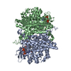
| Title | Crystal structure of the Acyl-CoA dehydrogenase FadE1(PA0506) E441A from Pseudomonas aeruginosa complexed with C16CoA |
|---|
 Components Components | Probable acyl-CoA dehydrogenase |
|---|
 Keywords Keywords | OXIDOREDUCTASE / acyl-CoA dehydrogenase / beta-oxidation |
|---|
| Function / homology |  Function and homology information Function and homology information
Acyl-CoA dehydrogenase, N-terminal, bacteria / : / Acetyl-CoA dehydrogenase-like C-terminal domain / Acetyl-CoA dehydrogenase C-terminal like / : / Acyl-CoA dehydrogenase/oxidase C-terminal / Acyl-CoA dehydrogenase/oxidase, N-terminal / Acyl-CoA dehydrogenase, N-terminal domain / Acyl-CoA dehydrogenase, C-terminal domain / Acyl-CoA oxidase/dehydrogenase, middle domain ...Acyl-CoA dehydrogenase, N-terminal, bacteria / : / Acetyl-CoA dehydrogenase-like C-terminal domain / Acetyl-CoA dehydrogenase C-terminal like / : / Acyl-CoA dehydrogenase/oxidase C-terminal / Acyl-CoA dehydrogenase/oxidase, N-terminal / Acyl-CoA dehydrogenase, N-terminal domain / Acyl-CoA dehydrogenase, C-terminal domain / Acyl-CoA oxidase/dehydrogenase, middle domain / Acyl-CoA dehydrogenase, middle domain / Acyl-CoA dehydrogenase/oxidase, N-terminal domain superfamily / Acyl-CoA oxidase/dehydrogenase, middle domain superfamily / Acyl-CoA dehydrogenase/oxidase, N-terminal and middle domain superfamily / Acyl-CoA dehydrogenase-like, C-terminalSimilarity search - Domain/homology |
|---|
| Biological species |   Pseudomonas aeruginosa (bacteria) Pseudomonas aeruginosa (bacteria) |
|---|
| Method |  X-RAY DIFFRACTION / SYNCHROTRON / MOLECULAR REPLACEMENT / Resolution: 1.44 Å X-RAY DIFFRACTION / SYNCHROTRON / MOLECULAR REPLACEMENT / Resolution: 1.44 Å |
|---|
 Authors Authors | Wang, M. / Brear, P. / Welch, M. |
|---|
| Funding support |  United Kingdom, 1items United Kingdom, 1items | Organization | Grant number | Country |
|---|
| Cystic Fibrosis Trust | SRC017 | United Kingdom |
|
|---|
 Citation Citation | Journal: Nat Commun / Year: 2025
Title: Pseudomonas aeruginosa acyl-CoA dehydrogenases and structure-guided inversion of their substrate specificity.
Authors: Wang, M. / Medarametla, P. / Kronenberger, T. / Deingruber, T. / Brear, P. / Figueroa, W. / Ho, P.M. / Krueger, T. / Pearce, J.C. / Poso, A. / Wakefield, J.G. / Spring, D.R. / Welch, M. |
|---|
| History | | Deposition | Jul 16, 2023 | Deposition site: PDBE / Processing site: PDBE |
|---|
| Revision 1.0 | Feb 12, 2025 | Provider: repository / Type: Initial release |
|---|
| Revision 1.1 | May 21, 2025 | Group: Database references / Category: citation / citation_author
Item: _citation.country / _citation.journal_abbrev ..._citation.country / _citation.journal_abbrev / _citation.journal_id_CSD / _citation.journal_id_ISSN / _citation.journal_volume / _citation.page_first / _citation.page_last / _citation.pdbx_database_id_DOI / _citation.pdbx_database_id_PubMed / _citation.title / _citation.year |
|---|
|
|---|
 Movie
Movie Controller

 Yorodumi
 Open data
 Basic information
 Structure visualization
 Downloads & links
 Other downloads
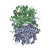

 PDBj
PDBj Assembly


 Mass: 1005.943 Da / Num. of mol.: 1 / Source method: obtained synthetically / Formula: C37H66N7O17P3S / Feature type: SUBJECT OF INVESTIGATION
 Mass: 62.068 Da / Num. of mol.: 4 / Source method: obtained synthetically / Formula: C2H6O2
 Mass: 150.173 Da / Num. of mol.: 1 / Source method: obtained synthetically / Formula: C6H14O4
 Mass: 62.005 Da / Num. of mol.: 2 / Source method: obtained synthetically / Formula: NO3
 Mass: 122.143 Da / Num. of mol.: 1 / Source method: obtained synthetically / Formula: C4H12NO3 / Comment: pH buffer*YM
 Sample preparation
 Processing