 Movie
Movie Controller


+ Open data

- Basic information
| Entry | Database: PDB / ID: 8ptt | ||||||
|---|---|---|---|---|---|---|---|
| Title | Human NUDT5 in complex with MRK-952 | ||||||
 Components Components | ADP-sugar pyrophosphatase | ||||||
 Keywords Keywords | HYDROLASE / Inhibitor | ||||||
| Function / homology |  Function and homology information Function and homology informationADP-D-ribose pyrophosphorylase / ribonucleoside diphosphate catabolic process / 8-oxo-dGDP phosphatase / ADP-sugar pyrophosphatase activity / nucleobase-containing small molecule metabolic process / ADP-ribose diphosphatase / D-ribose catabolic process / ADP-ribose diphosphatase activity / nucleoside phosphate metabolic process / 8-oxo-dGDP phosphatase activity ...ADP-D-ribose pyrophosphorylase / ribonucleoside diphosphate catabolic process / 8-oxo-dGDP phosphatase / ADP-sugar pyrophosphatase activity / nucleobase-containing small molecule metabolic process / ADP-ribose diphosphatase / D-ribose catabolic process / ADP-ribose diphosphatase activity / nucleoside phosphate metabolic process / 8-oxo-dGDP phosphatase activity / ribose phosphate metabolic process / Phosphate bond hydrolysis by NUDT proteins / ATP generation from poly-ADP-D-ribose / nucleotide metabolic process / snoRNA binding / nucleotidyltransferase activity / chromatin remodeling / magnesium ion binding / protein homodimerization activity / extracellular exosome / identical protein binding / nucleus / cytosol Similarity search - Function | ||||||
| Biological species |  Homo sapiens (human) Homo sapiens (human) | ||||||
| Method |  X-RAY DIFFRACTION / SYNCHROTRON / MOLECULAR REPLACEMENT / Resolution: 2.5 Å X-RAY DIFFRACTION / SYNCHROTRON / MOLECULAR REPLACEMENT / Resolution: 2.5 Å | ||||||
 Authors Authors | Diaz-Saez, L. / Koekemoer, L. / Feyerherm, C. / Sloman, S. / Fischer, C. / Schneider, S. / von Delft, F. / Arrowsmith, C.H. / Edwards, A.M. / Bountra, C. / Huber, K.V.M. | ||||||
| Funding support |  Canada, 1items Canada, 1items
| ||||||
 Citation Citation | Journal: To Be Published Title: Human NUDT5 in complex with MRK-952 Authors: Diaz-Saez, L. / Koekemoer, L. / Feyerherm, C. / Sloman, S. / Fischer, C. / Schneider, S. / von Delft, F. / Arrowsmith, C.H. / Edwards, A.M. / Bountra, C. / Huber, K.V.M. | ||||||
| History |
|
- Structure visualization
| Structure viewer | Molecule: MolmilJmol/JSmol |
|---|
- Downloads & links
-Download
| PDBx/mmCIF format | 8ptt.cif.gz | 180.6 KB | Display | PDBx/mmCIF format |
|---|---|---|---|---|
| PDB format | pdb8ptt.ent.gz | Display | PDB format | |
| PDBx/mmJSON format | 8ptt.json.gz | Tree view | PDBx/mmJSON format | |
| Others |  Other downloads Other downloads |
-Validation report
| Arichive directory | https://data.pdbj.org/pub/pdb/validation_reports/pt/8pttftp://data.pdbj.org/pub/pdb/validation_reports/pt/8ptt | HTTPS FTP |
|---|
-Related structure data
| Similar structure data |
|---|
-Links
 PDBj
PDBj

- Assembly
| Deposited unit | 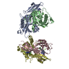
| ||||||||
|---|---|---|---|---|---|---|---|---|---|
| 1 | 
| ||||||||
| 2 | 
| ||||||||
| Unit cell |
|
-Components
| #1: Protein | Mass: 24355.596 Da / Num. of mol.: 4 Source method: isolated from a genetically manipulated source Source: (gene. exp.) Homo sapiens (human) / Gene: NUDT5, NUDIX5, HSPC115 / Production host:  References: UniProt: Q9UKK9, ADP-ribose diphosphatase, 8-oxo-dGDP phosphatase, ADP-D-ribose pyrophosphorylase #2: Chemical | ChemComp-F2V / Mass: 436.861 Da / Num. of mol.: 4 / Source method: obtained synthetically / Formula: C20H20ClF3N6 / Feature type: SUBJECT OF INVESTIGATION #3: Chemical | ChemComp-MG /   Mass: 24.305 Da / Num. of mol.: 7 / Source method: obtained synthetically / Formula: Mg Mass: 24.305 Da / Num. of mol.: 7 / Source method: obtained synthetically / Formula: Mg#4: Water | ChemComp-HOH / |  Mass: 18.015 Da / Num. of mol.: 290 / Source method: isolated from a natural source / Formula: H2O Mass: 18.015 Da / Num. of mol.: 290 / Source method: isolated from a natural source / Formula: H2OHas ligand of interest | Y | |
|---|
-Experimental details
-Experiment
| Experiment | Method: X-RAY DIFFRACTION / Number of used crystals: 1 |
|---|
- Sample preparation
| Crystal | Density Matthews: 2.32 Å3/Da / Density % sol: 46.95 % |
|---|---|
| Crystal grow | Temperature: 293 K / Method: vapor diffusion, sitting drop / pH: 8 Details: Reservoir: 33% PEG 400, 0.2 M magnesium chloride, and 0.1 M tris pH 8.0 |
-Data collection
| Diffraction | Mean temperature: 100 K / Serial crystal experiment: N |
|---|---|
| Diffraction source | Source: SYNCHROTRON / Site: Diamond  / Beamline: I03 / Wavelength: 0.97628 Å / Beamline: I03 / Wavelength: 0.97628 Å |
| Detector | Type: DECTRIS EIGER X 16M / Detector: PIXEL / Date: May 12, 2021 |
| Radiation | Protocol: SINGLE WAVELENGTH / Monochromatic (M) / Laue (L): M / Scattering type: x-ray |
| Radiation wavelength | Wavelength: 0.97628 Å / Relative weight: 1 |
| Reflection | Resolution: 2.5→28.41 Å / Num. obs: 29822 / % possible obs: 98.4 % / Redundancy: 3.2 % / Biso Wilson estimate: 33.048 Å2 / Rmerge(I) obs: 0.051 / Net I/σ(I): 9.2 |
| Reflection shell | Resolution: 2.5→2.6 Å / Redundancy: 3.5 % / Rmerge(I) obs: 0.225 / Mean I/σ(I) obs: 2.9 / Num. unique obs: 3405 / % possible all: 97.9 |
- Processing
| Software |
| |||||||||||||||||||||||||||||||||||||||||||||||||||||||||||||||||||||||||||||||||||||||||||||||||||||||||||||||||||||||||||||||||||||||||||||||||||||||||||
|---|---|---|---|---|---|---|---|---|---|---|---|---|---|---|---|---|---|---|---|---|---|---|---|---|---|---|---|---|---|---|---|---|---|---|---|---|---|---|---|---|---|---|---|---|---|---|---|---|---|---|---|---|---|---|---|---|---|---|---|---|---|---|---|---|---|---|---|---|---|---|---|---|---|---|---|---|---|---|---|---|---|---|---|---|---|---|---|---|---|---|---|---|---|---|---|---|---|---|---|---|---|---|---|---|---|---|---|---|---|---|---|---|---|---|---|---|---|---|---|---|---|---|---|---|---|---|---|---|---|---|---|---|---|---|---|---|---|---|---|---|---|---|---|---|---|---|---|---|---|---|---|---|---|---|---|---|
| Refinement | Method to determine structure: MOLECULAR REPLACEMENT / Resolution: 2.5→28.41 Å / Cor.coef. Fo:Fc: 0.956 / Cor.coef. Fo:Fc free: 0.917 / SU B: 10.15 / SU ML: 0.22 / Cross valid method: THROUGHOUT / ESU R: 0.815 / ESU R Free: 0.302
| |||||||||||||||||||||||||||||||||||||||||||||||||||||||||||||||||||||||||||||||||||||||||||||||||||||||||||||||||||||||||||||||||||||||||||||||||||||||||||
| Solvent computation | Ion probe radii: 0.8 Å / Shrinkage radii: 0.8 Å / VDW probe radii: 1.2 Å / Solvent model: MASK BULK SOLVENT | |||||||||||||||||||||||||||||||||||||||||||||||||||||||||||||||||||||||||||||||||||||||||||||||||||||||||||||||||||||||||||||||||||||||||||||||||||||||||||
| Displacement parameters | Biso mean: 39.036 Å2
| |||||||||||||||||||||||||||||||||||||||||||||||||||||||||||||||||||||||||||||||||||||||||||||||||||||||||||||||||||||||||||||||||||||||||||||||||||||||||||
| Refinement step | Cycle: LAST / Resolution: 2.5→28.41 Å
| |||||||||||||||||||||||||||||||||||||||||||||||||||||||||||||||||||||||||||||||||||||||||||||||||||||||||||||||||||||||||||||||||||||||||||||||||||||||||||
| Refine LS restraints |
| |||||||||||||||||||||||||||||||||||||||||||||||||||||||||||||||||||||||||||||||||||||||||||||||||||||||||||||||||||||||||||||||||||||||||||||||||||||||||||
| LS refinement shell |
|
