[English] 日本語
Yorodumi
- PDB-8por: Crystal structure of wolbachia leucyl-tRNA synthetase editing dom... -

+
Open data


ID or keywords:

Loading...

-
Basic information

Entry
Database: PDB / ID: 8por
TitleCrystal structure of wolbachia leucyl-tRNA synthetase editing domain bound to cmpd6-AMP adduct
ComponentsLeucine--tRNA ligase
KeywordsLIGASE / Leucine tRNA ligase / ATP binding protein / tRNA aminoacylation for protein translation / cytosolic Reaction catalysed: ATP + L-leucine + tRNA(Leu) = AMP + diphosphate + L-leucyl-tRNA(Leu)
Function / homology
Function and homology information


leucine-tRNA ligase / leucine-tRNA ligase activity / leucyl-tRNA aminoacylation / aminoacyl-tRNA editing activity / ATP binding / cytosol
Similarity search - Function
Leucyl-tRNA synthetase, editing domain / Leucyl-tRNA synthetase, editing domain / Leucine-tRNA ligase / Aminoacyl-tRNA synthetase, class Ia / tRNA synthetases class I (I, L, M and V) / Valyl/Leucyl/Isoleucyl-tRNA synthetase, editing domain / Methionyl/Valyl/Leucyl/Isoleucyl-tRNA synthetase, anticodon-binding / Anticodon-binding domain of tRNA ligase / Methionyl/Leucyl tRNA synthetase / tRNA synthetases class I (M) ...Leucyl-tRNA synthetase, editing domain / Leucyl-tRNA synthetase, editing domain / Leucine-tRNA ligase / Aminoacyl-tRNA synthetase, class Ia / tRNA synthetases class I (I, L, M and V) / Valyl/Leucyl/Isoleucyl-tRNA synthetase, editing domain / Methionyl/Valyl/Leucyl/Isoleucyl-tRNA synthetase, anticodon-binding / Anticodon-binding domain of tRNA ligase / Methionyl/Leucyl tRNA synthetase / tRNA synthetases class I (M) / Aminoacyl-tRNA synthetase, class Ia, anticodon-binding / Aminoacyl-tRNA synthetase, class I, conserved site / Aminoacyl-transfer RNA synthetases class-I signature. / Rossmann-like alpha/beta/alpha sandwich fold
Similarity search - Domain/homology
: / Leucine--tRNA ligase
Similarity search - Component
Biological speciesWolbachia endosymbiont strain TRS of Brugia malayi (bacteria)
MethodX-RAY DIFFRACTION / SYNCHROTRON / MOLECULAR REPLACEMENT / Resolution: 1.85 Å
AuthorsPalencia, A. / Lukarska, M.
Funding support France, European Union, 3items
OrganizationGrant numberCountry
Agence Nationale de la Recherche (ANR)ANR-20-AMRB-0003-01 AntibiOxaborole France
Agence Nationale de la Recherche (ANR)ANR JCJC RC18114CC NovoTargetParasite France
iNEXT653706European Union
CitationJournal: Science Advances / Year: 2024
Title: Targeting A Microbiota Wolbachian Aminoacyl-tRNA Synthetase To Block Its Pathogenic Host
Authors: Palencia, A. / Hoffmann, G. / Lukarska, M. / Clare, R. / Ward, S. / Taylor, M.A. / Ringkjobing Jensen, M.
History
DepositionJul 5, 2023Deposition site: PDBE / Processing site: PDBE
Revision 1.0Jun 12, 2024Provider: repository / Type: Initial release

-
Structure visualization

Structure viewerMolecule:
MolmilJmol/JSmol

Downloads & links

-
Assembly

Deposited unit
A: Leucine--tRNA ligase
hetero molecules


Theoretical massNumber of molelcules
Total (without water)21,2655
Polymers20,0121
Non-polymers1,2534
Water2,414134
1


  • Idetical with deposited unit
  • defined by author&software
  • Evidence: electron microscopy, elution volume suggests monomer, isothermal titration calorimetry, protein molecule (chain A) binds to 1 adduct cmpd6-AMP
TypeNameSymmetry operationNumber
identity operation1_555x,y,z1
Buried area620 Å2
ΔGint-4 kcal/mol
Surface area8830 Å2
MethodPISA
Unit cell
Length a, b, c (Å)126.630, 126.630, 61.590
Angle α, β, γ (deg.)90.00, 90.00, 120.00
Int Tables number178
Space group name H-MP6122
Components on special symmetry positions
IDModelComponents
11A-593-

HOH

21A-625-

HOH

-
Components

-
Protein , 1 types, 1 molecules A

#1: Protein Leucine--tRNA ligase / Leucyl-tRNA synthetase / LeuRS


Mass: 20012.182 Da / Num. of mol.: 1 / Mutation: deletion 354-477
Source method: isolated from a genetically manipulated source
Details: residue 30 (cysteine 243) is a S-mercaptocysteine (denoted CSS in PDB entries)
Source: (gene. exp.) Wolbachia endosymbiont strain TRS of Brugia malayi (bacteria)
Gene: leuS, Wbm0605 / Plasmid: pET-M11 / Production host: Escherichia coli BL21(DE3) (bacteria) / Variant (production host): RIL / References: UniProt: Q5GS31, leucine-tRNA ligase

-
Non-polymers , 5 types, 138 molecules

#2: Chemical ChemComp-SO4 / SULFATE ION


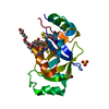
Mass: 96.063 Da / Num. of mol.: 1 / Source method: obtained synthetically / Formula: SO4
#3: Chemical ChemComp-EYT / [(1~{R},5~{S},6~{R},8~{R},9'~{S})-9'-(aminomethyl)-8-(6-aminopurin-9-yl)-2'-bromanyl-5'-[3-oxidanylidene-3-(1,3-thiazol-2-ylamino)propoxy]spiro[2,4,7-trioxa-3-boranuidabicyclo[3.3.0]octane-3,7'-8-oxa-7-boranuidabicyclo[4.3.0]nona-1(6),2,4-triene]-6-yl]methyl dihydrogen phosphate


Mass: 740.265 Da / Num. of mol.: 1 / Source method: obtained synthetically / Formula: C24H26BBrN8O10PS / Feature type: SUBJECT OF INVESTIGATION
#4: Chemical ChemComp-EDO / 1,2-ETHANEDIOL / ETHYLENE GLYCOL


Mass: 62.068 Da / Num. of mol.: 1 / Source method: obtained synthetically / Formula: C2H6O2
#5: Chemical ChemComp-PE4 / 2-{2-[2-(2-{2-[2-(2-ETHOXY-ETHOXY)-ETHOXY]-ETHOXY}-ETHOXY)-ETHOXY]-ETHOXY}-ETHANOL / POLYETHYLENE GLYCOL PEG4000


Mass: 354.436 Da / Num. of mol.: 1 / Source method: obtained synthetically / Formula: C16H34O8 / Comment: precipitant*YM
#6: Water ChemComp-HOH / water


Mass: 18.015 Da / Num. of mol.: 134 / Source method: isolated from a natural source / Formula: H2O

-
Details

Has ligand of interestY

-
Experimental details

-
Experiment

ExperimentMethod: X-RAY DIFFRACTION / Number of used crystals: 1

-
Sample preparation

CrystalDensity Matthews: 3.56 Å3/Da / Density % sol: 65.46 %
Crystal growTemperature: 293 K / Method: vapor diffusion, hanging drop / pH: 8.5 / Details: 0.1 M Tris pH 8.5; 0.2M LiSO4; 31% w/v PEG 4000

-
Data collection

DiffractionMean temperature: 100 K / Serial crystal experiment: N
Diffraction sourceSource: SYNCHROTRON / Site: ESRF / Beamline: ID23-1 / Wavelength: 0.97924 Å
DetectorType: DECTRIS PILATUS 6M / Detector: PIXEL / Date: Apr 18, 2014
RadiationProtocol: SINGLE WAVELENGTH / Monochromatic (M) / Laue (L): M / Scattering type: x-ray
Radiation wavelengthWavelength: 0.97924 Å / Relative weight: 1
ReflectionResolution: 1.85→109.66 Å / Num. obs: 24048 / % possible obs: 100 % / Redundancy: 16.2 % / CC1/2: 1 / Rrim(I) all: 0.09 / Net I/σ(I): 21.91
Reflection shellResolution: 1.85→1.93 Å / Redundancy: 16.8 % / Mean I/σ(I) obs: 1.62 / Num. unique obs: 1281 / CC1/2: 0.67 / Rrim(I) all: 1.89 / % possible all: 100

-
Processing

Software
NameVersionClassification
REFMAC5.8.0258refinement
XDSdata reduction
XSCALEdata scaling
PHASERphasing
RefinementMethod to determine structure: MOLECULAR REPLACEMENT / Resolution: 1.85→44.19 Å / Cor.coef. Fo:Fc: 0.975 / Cor.coef. Fo:Fc free: 0.975 / SU B: 6.577 / SU ML: 0.08 / Cross valid method: THROUGHOUT / ESU R: 0.107 / ESU R Free: 0.079 / Stereochemistry target values: MAXIMUM LIKELIHOOD / Details: HYDROGENS HAVE BEEN ADDED IN THE RIDING POSITIONS
RfactorNum. reflection% reflectionSelection details
Rfree0.16602 1281 5.1 %RANDOM
Rwork0.1471 ---
obs0.14812 24048 99.98 %-
Solvent computationIon probe radii: 0.8 Å / Shrinkage radii: 0.8 Å / VDW probe radii: 1.2 Å / Solvent model: MASK
Displacement parametersBiso mean: 37.348 Å2
Baniso -1Baniso -2Baniso -3
1--2.06 Å2-1.03 Å20 Å2
2---2.06 Å20 Å2
3---6.68 Å2
Refinement stepCycle: 1 / Resolution: 1.85→44.19 Å
ProteinNucleic acidLigandSolventTotal
Num. atoms1293 0 77 134 1504
Refine LS restraints
Refine-IDTypeDev idealDev ideal targetNumber
X-RAY DIFFRACTIONr_bond_refined_d0.0060.0131433
X-RAY DIFFRACTIONr_bond_other_d0.0030.0171366
X-RAY DIFFRACTIONr_angle_refined_deg1.8691.7061943
X-RAY DIFFRACTIONr_angle_other_deg1.2961.6283192
X-RAY DIFFRACTIONr_dihedral_angle_1_deg7.165174
X-RAY DIFFRACTIONr_dihedral_angle_2_deg29.6192562
X-RAY DIFFRACTIONr_dihedral_angle_3_deg14.42115252
X-RAY DIFFRACTIONr_dihedral_angle_4_deg14.181153
X-RAY DIFFRACTIONr_chiral_restr0.2190.2192
X-RAY DIFFRACTIONr_gen_planes_refined0.0050.021519
X-RAY DIFFRACTIONr_gen_planes_other0.0020.02257
X-RAY DIFFRACTIONr_nbd_refined
X-RAY DIFFRACTIONr_nbd_other
X-RAY DIFFRACTIONr_nbtor_refined
X-RAY DIFFRACTIONr_nbtor_other
X-RAY DIFFRACTIONr_xyhbond_nbd_refined
X-RAY DIFFRACTIONr_xyhbond_nbd_other
X-RAY DIFFRACTIONr_metal_ion_refined
X-RAY DIFFRACTIONr_metal_ion_other
X-RAY DIFFRACTIONr_symmetry_vdw_refined
X-RAY DIFFRACTIONr_symmetry_vdw_other
X-RAY DIFFRACTIONr_symmetry_hbond_refined
X-RAY DIFFRACTIONr_symmetry_hbond_other
X-RAY DIFFRACTIONr_symmetry_metal_ion_refined
X-RAY DIFFRACTIONr_symmetry_metal_ion_other
X-RAY DIFFRACTIONr_mcbond_it3.8324.532666
X-RAY DIFFRACTIONr_mcbond_other3.82615.944665
X-RAY DIFFRACTIONr_mcangle_it5.143832
X-RAY DIFFRACTIONr_mcangle_other5.169833
X-RAY DIFFRACTIONr_scbond_it5.0753.958766
X-RAY DIFFRACTIONr_scbond_other5.072767
X-RAY DIFFRACTIONr_scangle_it
X-RAY DIFFRACTIONr_scangle_other6.43145.9911106
X-RAY DIFFRACTIONr_long_range_B_refined7.6921528
X-RAY DIFFRACTIONr_long_range_B_other7.5761507
X-RAY DIFFRACTIONr_rigid_bond_restr3.05531427
X-RAY DIFFRACTIONr_sphericity_free
X-RAY DIFFRACTIONr_sphericity_bonded
LS refinement shellResolution: 1.85→1.898 Å / Total num. of bins used: 20
RfactorNum. reflection% reflection
Rfree0.318 115 -
Rwork0.303 1717 -
obs--100 %

+
About Yorodumi

-
News

-
Feb 9, 2022. New format data for meta-information of EMDB entries

New format data for meta-information of EMDB entries

  • Version 3 of the EMDB header file is now the official format.
  • The previous official version 1.9 will be removed from the archive.

Related info.:EMDB header

External links:wwPDB to switch to version 3 of the EMDB data model

-
Aug 12, 2020. Covid-19 info

Covid-19 info

URL: https://pdbjlvh1.pdbj.org/emnavi/covid19.php

New page: Covid-19 featured information page in EM Navigator.

Related info.:Covid-19 info / Mar 5, 2020. Novel coronavirus structure data

+
Mar 5, 2020. Novel coronavirus structure data

Novel coronavirus structure data

Related info.:Yorodumi Speices / Aug 12, 2020. Covid-19 info

External links:COVID-19 featured content - PDBj / Molecule of the Month (242):Coronavirus Proteases

+
Jan 31, 2019. EMDB accession codes are about to change! (news from PDBe EMDB page)

EMDB accession codes are about to change! (news from PDBe EMDB page)

  • The allocation of 4 digits for EMDB accession codes will soon come to an end. Whilst these codes will remain in use, new EMDB accession codes will include an additional digit and will expand incrementally as the available range of codes is exhausted. The current 4-digit format prefixed with “EMD-” (i.e. EMD-XXXX) will advance to a 5-digit format (i.e. EMD-XXXXX), and so on. It is currently estimated that the 4-digit codes will be depleted around Spring 2019, at which point the 5-digit format will come into force.
  • The EM Navigator/Yorodumi systems omit the EMD- prefix.

Related info.:Q: What is EMD? / ID/Accession-code notation in Yorodumi/EM Navigator

External links:EMDB Accession Codes are Changing Soon! / Contact to PDBj

+
Jul 12, 2017. Major update of PDB

Major update of PDB

  • wwPDB released updated PDB data conforming to the new PDBx/mmCIF dictionary.
  • This is a major update changing the version number from 4 to 5, and with Remediation, in which all the entries are updated.
  • In this update, many items about electron microscopy experimental information are reorganized (e.g. em_software).
  • Now, EM Navigator and Yorodumi are based on the updated data.

External links:wwPDB Remediation / Enriched Model Files Conforming to OneDep Data Standards Now Available in the PDB FTP Archive

-
Yorodumi

Thousand views of thousand structures

  • Yorodumi is a browser for structure data from EMDB, PDB, SASBDB, etc.
  • This page is also the successor to EM Navigator detail page, and also detail information page/front-end page for Omokage search.
  • The word "yorodu" (or yorozu) is an old Japanese word meaning "ten thousand". "mi" (miru) is to see.

Related info.:EMDB / PDB / SASBDB / Comparison of 3 databanks / Yorodumi Search / Aug 31, 2016. New EM Navigator & Yorodumi / Yorodumi Papers / Jmol/JSmol / Function and homology information / Changes in new EM Navigator and Yorodumi

Read more