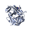
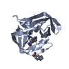
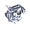
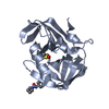
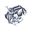
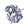
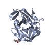
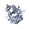
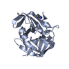
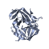
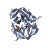
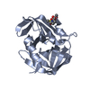
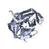
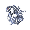
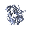
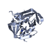
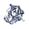
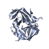
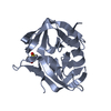
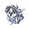
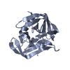
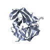
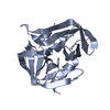
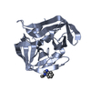
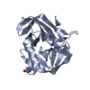
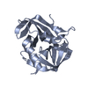
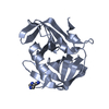
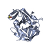
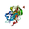
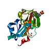
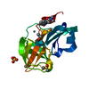
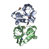
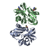
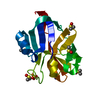
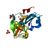
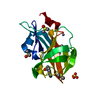
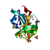
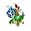
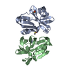
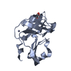
[English] 日本語
Yorodumi
- PDB-8poa: Structure of Coxsackievirus A16 (G-10) 2A protease -

+
Open data


ID or keywords:

Loading...

-
Basic information

Entry
Database: PDB / ID: 8poa
TitleStructure of Coxsackievirus A16 (G-10) 2A protease
ComponentsProtease 2A
KeywordsVIRAL PROTEIN / Enterovirus 2A protease ASAP AViDD Cysteine / Cysteine protease / ANTIVIRAL PROTEIN
Function / homology
Function and homology information


symbiont-mediated suppression of host cytoplasmic pattern recognition receptor signaling pathway via inhibition of MDA-5 activity / picornain 2A / symbiont-mediated suppression of host mRNA export from nucleus / symbiont genome entry into host cell via pore formation in plasma membrane / picornain 3C / T=pseudo3 icosahedral viral capsid / host cell cytoplasmic vesicle membrane / endocytosis involved in viral entry into host cell / : / nucleoside-triphosphate phosphatase ...symbiont-mediated suppression of host cytoplasmic pattern recognition receptor signaling pathway via inhibition of MDA-5 activity / picornain 2A / symbiont-mediated suppression of host mRNA export from nucleus / symbiont genome entry into host cell via pore formation in plasma membrane / picornain 3C / T=pseudo3 icosahedral viral capsid / host cell cytoplasmic vesicle membrane / endocytosis involved in viral entry into host cell / : / nucleoside-triphosphate phosphatase / protein complex oligomerization / monoatomic ion channel activity / : / RNA helicase activity / DNA replication / induction by virus of host autophagy / RNA-directed RNA polymerase / symbiont-mediated suppression of host gene expression / viral RNA genome replication / cysteine-type endopeptidase activity / RNA-dependent RNA polymerase activity / DNA-templated transcription / host cell nucleus / virion attachment to host cell / structural molecule activity / ATP hydrolysis activity / proteolysis / RNA binding / ATP binding / membrane / metal ion binding
Similarity search - Function
Poliovirus 3A protein-like / Poliovirus 3A protein like / Picornavirus 2B protein / Poliovirus core protein 3a, soluble domain / Picornavirus 2B protein / Peptidase C3, picornavirus core protein 2A / Picornavirus core protein 2A / Picornavirus coat protein VP4 / Picornavirus coat protein (VP4) / Picornavirales 3C/3C-like protease domain ...Poliovirus 3A protein-like / Poliovirus 3A protein like / Picornavirus 2B protein / Poliovirus core protein 3a, soluble domain / Picornavirus 2B protein / Peptidase C3, picornavirus core protein 2A / Picornavirus core protein 2A / Picornavirus coat protein VP4 / Picornavirus coat protein (VP4) / Picornavirales 3C/3C-like protease domain / Picornavirales 3C/3C-like protease domain profile. / Peptidase C3A/C3B, picornaviral / 3C cysteine protease (picornain 3C) / Picornavirus capsid / picornavirus capsid protein / Helicase, superfamily 3, single-stranded RNA virus / Superfamily 3 helicase of positive ssRNA viruses domain profile. / Helicase, superfamily 3, single-stranded DNA/RNA virus / RNA helicase / Picornavirus/Calicivirus coat protein / Viral coat protein subunit / RNA-directed RNA polymerase, C-terminal domain / Viral RNA-dependent RNA polymerase / Reverse transcriptase/Diguanylate cyclase domain / RNA-directed RNA polymerase, catalytic domain / RdRp of positive ssRNA viruses catalytic domain profile. / ATPases associated with a variety of cellular activities / AAA+ ATPase domain / Peptidase S1, PA clan, chymotrypsin-like fold / Peptidase S1, PA clan / DNA/RNA polymerase superfamily / P-loop containing nucleoside triphosphate hydrolase
Similarity search - Domain/homology
Biological speciesCoxsackievirus A16
MethodX-RAY DIFFRACTION / SYNCHROTRON / MOLECULAR REPLACEMENT / Resolution: 1.6 Å
AuthorsLithgo, R.M. / Fairhead, M. / Koekemoer, L. / Aschenbrenner, J.C. / Balcomb, B.H. / Godoy, A.S. / Marples, P.G. / Ni, X. / Tomlinson, C.W.E. / Thompson, W. ...Lithgo, R.M. / Fairhead, M. / Koekemoer, L. / Aschenbrenner, J.C. / Balcomb, B.H. / Godoy, A.S. / Marples, P.G. / Ni, X. / Tomlinson, C.W.E. / Thompson, W. / Wild, C. / Fearon, D. / Walsh, M.A. / von Delft, F.
Funding support United States, 1items
OrganizationGrant numberCountry
National Institutes of Health/National Institute Of Allergy and Infectious Diseases (NIH/NIAID)U19AI71399 United States
CitationJournal: To Be Published
Title: Structure of EV A71 2A protease - to be published
Authors: Lithgo, R.M. / von Delft, F.
History
DepositionJul 4, 2023Deposition site: PDBE / Processing site: PDBE
Revision 1.0Aug 2, 2023Provider: repository / Type: Initial release
Revision 1.1Mar 20, 2024Group: Data collection / Derived calculations ...Data collection / Derived calculations / Source and taxonomy / Structure summary
Category: chem_comp_atom / chem_comp_bond ...chem_comp_atom / chem_comp_bond / entity / entity_name_com / entity_src_gen / pdbx_struct_assembly / pdbx_struct_assembly_gen / struct
Item: _entity.pdbx_description / _entity.pdbx_ec ..._entity.pdbx_description / _entity.pdbx_ec / _entity_name_com.name / _entity_src_gen.pdbx_gene_src_ncbi_taxonomy_id / _entity_src_gen.pdbx_gene_src_scientific_name / _struct.title

-
Structure visualization

Structure viewerMolecule:
MolmilJmol/JSmol

Downloads & links

-
Assembly

Deposited unit
A: Protease 2A
B: Protease 2A
hetero molecules


Theoretical massNumber of molelcules
Total (without water)32,1006
Polymers31,7852
Non-polymers3154
Water6,630368
1
A: Protease 2A
hetero molecules


Theoretical massNumber of molelcules
Total (without water)16,0503
Polymers15,8931
Non-polymers1582
Water181
TypeNameSymmetry operationNumber
identity operation1_555x,y,z1
2
B: Protease 2A
hetero molecules


Theoretical massNumber of molelcules
Total (without water)16,0503
Polymers15,8931
Non-polymers1582
Water181
TypeNameSymmetry operationNumber
identity operation1_555x,y,z1
Unit cell
Length a, b, c (Å)86.128, 57.199, 64.631
Angle α, β, γ (deg.)90.00, 95.23, 90.00
Int Tables number5
Space group name H-MC121

-
Components

#1: Protein Protease 2A / P2A / Picornain 2A / Protein 2A


Mass: 15892.732 Da / Num. of mol.: 2
Source method: isolated from a genetically manipulated source
Source: (gene. exp.) Coxsackievirus A16 (strain G-10) / Production host: Escherichia coli BL21 (bacteria) / References: UniProt: Q65900, picornain 2A
#2: Chemical ChemComp-GOL / GLYCEROL / GLYCERIN / PROPANE-1,2,3-TRIOL / Glycerol


Mass: 92.094 Da / Num. of mol.: 2 / Source method: obtained synthetically / Formula: C3H8O3
#3: Chemical ChemComp-ZN / ZINC ION


Mass: 65.409 Da / Num. of mol.: 2 / Source method: obtained synthetically / Formula: Zn / Feature type: SUBJECT OF INVESTIGATION
#4: Water ChemComp-HOH / water / Water


Mass: 18.015 Da / Num. of mol.: 368 / Source method: isolated from a natural source / Formula: H2O
Has ligand of interestY

-
Experimental details

-
Experiment

ExperimentMethod: X-RAY DIFFRACTION / Number of used crystals: 1

-
Sample preparation

CrystalDensity Matthews: 2.49 Å3/Da / Density % sol: 50.68 %
Crystal growTemperature: 298 K / Method: vapor diffusion, sitting drop / Details: 0.2M Ammonium Formate 20% PEG 3350

-
Data collection

DiffractionMean temperature: 100 K / Serial crystal experiment: N
Diffraction sourceSource: SYNCHROTRON / Site: Diamond / Beamline: I04-1 / Wavelength: 0.9212 Å
DetectorType: DECTRIS EIGER2 X 9M / Detector: PIXEL / Date: May 22, 2023
RadiationProtocol: SINGLE WAVELENGTH / Monochromatic (M) / Laue (L): M / Scattering type: x-ray
Radiation wavelengthWavelength: 0.9212 Å / Relative weight: 1
ReflectionResolution: 1.6→16.09 Å / Num. obs: 41210 / % possible obs: 99.5 % / Redundancy: 6.7 % / Biso Wilson estimate: 15.18 Å2 / CC1/2: 0.996 / CC star: 0.999 / Rmerge(I) obs: 0.1461 / Rpim(I) all: 0.05993 / Rrim(I) all: 0.1581 / Net I/σ(I): 10.43
Reflection shellResolution: 1.6→1.657 Å / Redundancy: 5.3 % / Rmerge(I) obs: 0.7042 / Mean I/σ(I) obs: 1.63 / Num. unique obs: 4072 / CC1/2: 0.653 / CC star: 0.889 / Rpim(I) all: 0.3338 / Rrim(I) all: 0.7818 / % possible all: 98.91

-
Processing

Software
NameVersionClassification
BUSTER2.10.4 (14-JUN-2023)refinement
autoPROCdata reduction
autoPROCdata scaling
PHASERphasing
RefinementMethod to determine structure: MOLECULAR REPLACEMENT / Resolution: 1.6→16.09 Å / Cor.coef. Fo:Fc: 0.957 / Cor.coef. Fo:Fc free: 0.952 / SU R Cruickshank DPI: 0.086 / Cross valid method: THROUGHOUT / σ(F): 0 / SU R Blow DPI: 0.093 / SU Rfree Blow DPI: 0.088 / SU Rfree Cruickshank DPI: 0.084
RfactorNum. reflection% reflectionSelection details
Rfree0.2098 2015 4.89 %RANDOM
Rwork0.1858 ---
obs0.187 41210 99.6 %-
Displacement parametersBiso mean: 18.81 Å2
Baniso -1Baniso -2Baniso -3
1--0.2811 Å20 Å2-1.2878 Å2
2---0.6548 Å20 Å2
3---0.9359 Å2
Refine analyzeLuzzati coordinate error obs: 0.2 Å
Refinement stepCycle: LAST / Resolution: 1.6→16.09 Å
ProteinNucleic acidLigandSolventTotal
Num. atoms2166 0 14 368 2548
Refine LS restraints
Refine-IDTypeDev idealNumberRestraint functionWeight
X-RAY DIFFRACTIONt_bond_d0.012266HARMONIC2
X-RAY DIFFRACTIONt_angle_deg0.933085HARMONIC2
X-RAY DIFFRACTIONt_dihedral_angle_d760SINUSOIDAL2
X-RAY DIFFRACTIONt_incorr_chiral_ct
X-RAY DIFFRACTIONt_pseud_angle
X-RAY DIFFRACTIONt_trig_c_planes
X-RAY DIFFRACTIONt_gen_planes397HARMONIC5
X-RAY DIFFRACTIONt_it2266HARMONIC10
X-RAY DIFFRACTIONt_nbd
X-RAY DIFFRACTIONt_omega_torsion4.13
X-RAY DIFFRACTIONt_other_torsion15.06
X-RAY DIFFRACTIONt_improper_torsion
X-RAY DIFFRACTIONt_chiral_improper_torsion279SEMIHARMONIC5
X-RAY DIFFRACTIONt_sum_occupancies
X-RAY DIFFRACTIONt_utility_distance
X-RAY DIFFRACTIONt_utility_angle
X-RAY DIFFRACTIONt_utility_torsion
X-RAY DIFFRACTIONt_ideal_dist_contact2156SEMIHARMONIC4
LS refinement shellResolution: 1.6→1.61 Å / Total num. of bins used: 51
RfactorNum. reflection% reflection
Rfree0.3014 -3.88 %
Rwork0.2546 793 -
all0.2564 825 -
obs--98.45 %
Refinement TLS params.

T22: -0.0148 Å2 / Method: refined / Refine-ID: X-RAY DIFFRACTION

IDL112)L122)L132)L222)L232)L332)S11 (Å °)S12 (Å °)S13 (Å °)S21 (Å °)S22 (Å °)S23 (Å °)S31 (Å °)S32 (Å °)S33 (Å °)T112)T122)T132)T232)T332)Origin x (Å)Origin y (Å)Origin z (Å)
10.5730.0972-0.01120.7834-0.00520.28160.0057-0.00160.085-0.00730.038-0.03920.0372-0.0236-0.0436-0.021-0.0039-0.0018-0.0115-0.008114.54177.327114.8677
20.7806-0.2919-0.13440.4723-0.08610.42190.0035-0.02090.059-0.00990.00880.02090.02160.0307-0.0123-0.0195-0.01020.0009-0.0014-0.009525.255135.574117.895
Refinement TLS group
IDRefine-IDRefine TLS-IDSelection details
1X-RAY DIFFRACTION1{ A|* }
2X-RAY DIFFRACTION2{ B|* }

+
About Yorodumi

-
News

-
Feb 9, 2022. New format data for meta-information of EMDB entries

New format data for meta-information of EMDB entries

  • Version 3 of the EMDB header file is now the official format.
  • The previous official version 1.9 will be removed from the archive.

Related info.:EMDB header

External links:wwPDB to switch to version 3 of the EMDB data model

-
Aug 12, 2020. Covid-19 info

Covid-19 info

URL: https://pdbj.org/emnavi/covid19.php

New page: Covid-19 featured information page in EM Navigator.

Related info.:Covid-19 info / Mar 5, 2020. Novel coronavirus structure data

+
Mar 5, 2020. Novel coronavirus structure data

Novel coronavirus structure data

Related info.:Yorodumi Speices / Aug 12, 2020. Covid-19 info

External links:COVID-19 featured content - PDBj / Molecule of the Month (242):Coronavirus Proteases

+
Jan 31, 2019. EMDB accession codes are about to change! (news from PDBe EMDB page)

EMDB accession codes are about to change! (news from PDBe EMDB page)

  • The allocation of 4 digits for EMDB accession codes will soon come to an end. Whilst these codes will remain in use, new EMDB accession codes will include an additional digit and will expand incrementally as the available range of codes is exhausted. The current 4-digit format prefixed with “EMD-” (i.e. EMD-XXXX) will advance to a 5-digit format (i.e. EMD-XXXXX), and so on. It is currently estimated that the 4-digit codes will be depleted around Spring 2019, at which point the 5-digit format will come into force.
  • The EM Navigator/Yorodumi systems omit the EMD- prefix.

Related info.:Q: What is EMD? / ID/Accession-code notation in Yorodumi/EM Navigator

External links:EMDB Accession Codes are Changing Soon! / Contact to PDBj

+
Jul 12, 2017. Major update of PDB

Major update of PDB

  • wwPDB released updated PDB data conforming to the new PDBx/mmCIF dictionary.
  • This is a major update changing the version number from 4 to 5, and with Remediation, in which all the entries are updated.
  • In this update, many items about electron microscopy experimental information are reorganized (e.g. em_software).
  • Now, EM Navigator and Yorodumi are based on the updated data.

External links:wwPDB Remediation / Enriched Model Files Conforming to OneDep Data Standards Now Available in the PDB FTP Archive

-
Yorodumi

Thousand views of thousand structures

  • Yorodumi is a browser for structure data from EMDB, PDB, SASBDB, etc.
  • This page is also the successor to EM Navigator detail page, and also detail information page/front-end page for Omokage search.
  • The word "yorodu" (or yorozu) is an old Japanese word meaning "ten thousand". "mi" (miru) is to see.

Related info.:EMDB / PDB / SASBDB / Comparison of 3 databanks / Yorodumi Search / Aug 31, 2016. New EM Navigator & Yorodumi / Yorodumi Papers / Jmol/JSmol / Function and homology information / Changes in new EM Navigator and Yorodumi

Read more