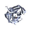
[English] 日本語
Yorodumi
- PDB-7gpr: PanDDA analysis group deposition -- Crystal Structure of Enterovi... -

+
Open data


ID or keywords:

Loading...

-
Basic information

Entry
Database: PDB / ID: 7gpr
TitlePanDDA analysis group deposition -- Crystal Structure of Enterovirus D68 3C Protease in complex with POB0095
ComponentsProtease 3CPicornain 3C
KeywordsVIRAL PROTEIN / Diamond I04-1 fragment screening / PanDDA / XChemExplorer / XCE / Viral Protease / 3C
Function / homology
Function and homology information


symbiont-mediated suppression of cytoplasmic pattern recognition receptor signaling pathway / symbiont-mediated suppression of host cytoplasmic pattern recognition receptor signaling pathway via inhibition of MDA-5 activity / picornain 2A / symbiont-mediated suppression of host mRNA export from nucleus / symbiont genome entry into host cell via pore formation in plasma membrane / protein sequestering activity / picornain 3C / T=pseudo3 icosahedral viral capsid / host cell cytoplasmic vesicle membrane / endocytosis involved in viral entry into host cell ...symbiont-mediated suppression of cytoplasmic pattern recognition receptor signaling pathway / symbiont-mediated suppression of host cytoplasmic pattern recognition receptor signaling pathway via inhibition of MDA-5 activity / picornain 2A / symbiont-mediated suppression of host mRNA export from nucleus / symbiont genome entry into host cell via pore formation in plasma membrane / protein sequestering activity / picornain 3C / T=pseudo3 icosahedral viral capsid / host cell cytoplasmic vesicle membrane / endocytosis involved in viral entry into host cell / : / nucleoside-triphosphate phosphatase / protein complex oligomerization / monoatomic ion channel activity / : / symbiont-mediated suppression of host toll-like receptor signaling pathway / RNA helicase activity / induction by virus of host autophagy / RNA-directed RNA polymerase / symbiont-mediated suppression of host gene expression / viral RNA genome replication / cysteine-type endopeptidase activity / RNA-dependent RNA polymerase activity / DNA-templated transcription / host cell nucleus / virion attachment to host cell / structural molecule activity / ATP hydrolysis activity / proteolysis / RNA binding / ATP binding / membrane / metal ion binding / cytoplasm
Similarity search - Function
Poliovirus 3A protein-like / Poliovirus 3A protein like / Picornavirus 2B protein / Poliovirus core protein 3a, soluble domain / Picornavirus 2B protein / Peptidase C3, picornavirus core protein 2A / Picornavirus core protein 2A / Picornavirus coat protein VP4 / Picornavirus coat protein (VP4) / Picornavirales 3C/3C-like protease domain ...Poliovirus 3A protein-like / Poliovirus 3A protein like / Picornavirus 2B protein / Poliovirus core protein 3a, soluble domain / Picornavirus 2B protein / Peptidase C3, picornavirus core protein 2A / Picornavirus core protein 2A / Picornavirus coat protein VP4 / Picornavirus coat protein (VP4) / Picornavirales 3C/3C-like protease domain / Picornavirales 3C/3C-like protease domain profile. / Peptidase C3A/C3B, picornaviral / 3C cysteine protease (picornain 3C) / Picornavirus capsid / picornavirus capsid protein / Helicase, superfamily 3, single-stranded RNA virus / Superfamily 3 helicase of positive ssRNA viruses domain profile. / Helicase, superfamily 3, single-stranded DNA/RNA virus / RNA helicase / Picornavirus/Calicivirus coat protein / Viral coat protein subunit / RNA-directed RNA polymerase, C-terminal domain / Viral RNA-dependent RNA polymerase / Reverse transcriptase/Diguanylate cyclase domain / RNA-directed RNA polymerase, catalytic domain / RdRp of positive ssRNA viruses catalytic domain profile. / ATPases associated with a variety of cellular activities / AAA+ ATPase domain / Peptidase S1, PA clan, chymotrypsin-like fold / Peptidase S1, PA clan / DNA/RNA polymerase superfamily / P-loop containing nucleoside triphosphate hydrolase
Similarity search - Domain/homology
Chem-U1I / Genome polyprotein
Similarity search - Component
Biological speciesHuman Enterovirus D68
MethodX-RAY DIFFRACTION / SYNCHROTRON / MOLECULAR REPLACEMENT / Resolution: 1.65 Å
AuthorsLithgo, R.M. / Fairhead, M. / Koekemoer, L. / Aschenbrenner, J.C. / Balcomb, B.H. / Godoy, A.S. / Marples, P.G. / Ni, X. / Tomlinson, C.W.E. / Thompson, W. ...Lithgo, R.M. / Fairhead, M. / Koekemoer, L. / Aschenbrenner, J.C. / Balcomb, B.H. / Godoy, A.S. / Marples, P.G. / Ni, X. / Tomlinson, C.W.E. / Thompson, W. / Wild, C. / Fearon, D. / Walsh, M.A. / von Delft, F.
Funding support United States, 1items
OrganizationGrant numberCountry
National Institutes of Health/National Institute Of Allergy and Infectious Diseases (NIH/NIAID)1U19AI171399 United States
CitationJournal: To Be Published
Title: PanDDA analysis group deposition
Authors: Lithgo, R.M. / Fairhead, M. / Koekemoer, L. / Aschenbrenner, J.C. / Balcomb, B.H. / Godoy, A.S. / Marples, P.G. / Ni, X. / Tomlinson, C.W.E. / Thompson, W. / Wild, C. / Fearon, D. / Walsh, M.A. / von Delft, F.
History
DepositionAug 24, 2023Deposition site: RCSB / Processing site: RCSB
Revision 1.0Nov 29, 2023Provider: repository / Type: Initial release

-
Structure visualization

Structure viewerMolecule:
MolmilJmol/JSmol

Downloads & links

-
Assembly

Deposited unit
A: Protease 3C
B: Protease 3C
hetero molecules


Theoretical massNumber of molelcules
Total (without water)40,5494
Polymers40,2522
Non-polymers2972
Water5,621312
1
A: Protease 3C
hetero molecules


Theoretical massNumber of molelcules
Total (without water)20,3452
Polymers20,1261
Non-polymers2191
Water181
TypeNameSymmetry operationNumber
identity operation1_555x,y,z1
2
B: Protease 3C
hetero molecules


Theoretical massNumber of molelcules
Total (without water)20,2042
Polymers20,1261
Non-polymers781
Water181
TypeNameSymmetry operationNumber
identity operation1_555x,y,z1
Unit cell
Length a, b, c (Å)42.726, 62.417, 148.217
Angle α, β, γ (deg.)90.00, 90.00, 90.00
Int Tables number19
Space group name H-MP212121

-
Components

#1: Protein Protease 3C / Picornain 3C / Picornain 3C / P3C


Mass: 20126.010 Da / Num. of mol.: 2
Source method: isolated from a genetically manipulated source
Source: (gene. exp.) Human Enterovirus D68 / Production host: Escherichia coli (E. coli) / References: UniProt: Q68T42, picornain 3C
#2: Chemical ChemComp-U1I / methyl (3R)-3-(4-methylphenyl)pyrrolidine-3-carboxylate


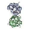
Mass: 219.280 Da / Num. of mol.: 1 / Source method: obtained synthetically / Formula: C13H17NO2 / Feature type: SUBJECT OF INVESTIGATION
#3: Chemical ChemComp-DMS / DIMETHYL SULFOXIDE / Dimethyl sulfoxide


Mass: 78.133 Da / Num. of mol.: 1 / Source method: obtained synthetically / Formula: C2H6OS / Comment: DMSO, precipitant*YM
#4: Water ChemComp-HOH / water / Water


Mass: 18.015 Da / Num. of mol.: 312 / Source method: isolated from a natural source / Formula: H2O
Has ligand of interestY

-
Experimental details

-
Experiment

ExperimentMethod: X-RAY DIFFRACTION / Number of used crystals: 1

-
Sample preparation

CrystalDensity Matthews: 2.45 Å3/Da / Density % sol: 49.9 %
Crystal growTemperature: 298 K / Method: vapor diffusion, sitting drop / pH: 8.14 / Details: 25% PEG 3350, 0.1M Tris, 0.2M Ammonium acetate

-
Data collection

DiffractionMean temperature: 100 K
Diffraction sourceSource: SYNCHROTRON / Site: Diamond / Beamline: I04-1 / Wavelength: 0.92124 Å
DetectorType: DECTRIS PILATUS 6M / Detector: PIXEL / Date: Jan 26, 2023
RadiationProtocol: SINGLE WAVELENGTH / Scattering type: x-ray
Radiation wavelengthWavelength: 0.92124 Å / Relative weight: 1
ReflectionResolution: 1.65→38.74 Å / Num. obs: 48702 / % possible obs: 99.8 % / Redundancy: 13.4 % / CC1/2: 0.997 / Rmerge(I) obs: 0.232 / Rpim(I) all: 0.066 / Rrim(I) all: 0.241 / Χ2: 0.41 / Net I/σ(I): 11.1 / Num. measured all: 653482
Reflection shellResolution: 1.65→1.68 Å / % possible obs: 96.4 % / Redundancy: 13.8 % / Rmerge(I) obs: 5.68 / Num. measured all: 31517 / Num. unique obs: 2286 / CC1/2: 0.299 / Rpim(I) all: 1.592 / Rrim(I) all: 5.902 / Χ2: 0.12 / Net I/σ(I) obs: 0.9

-
Processing

Software
NameVersionClassification
REFMAC5.8.0267refinement
Aimlessdata scaling
PHASERphasing
XDSdata reduction
RefinementMethod to determine structure: MOLECULAR REPLACEMENT / Resolution: 1.65→38.77 Å / Cor.coef. Fo:Fc: 0.965 / Cor.coef. Fo:Fc free: 0.949 / SU B: 2.935 / SU ML: 0.092 / Cross valid method: THROUGHOUT / ESU R: 0.106 / ESU R Free: 0.106 / Stereochemistry target values: MAXIMUM LIKELIHOOD / Details: HYDROGENS HAVE BEEN ADDED IN THE RIDING POSITIONS
RfactorNum. reflection% reflectionSelection details
Rfree0.22121 2419 5 %RANDOM
Rwork0.18362 ---
obs0.18545 45603 98.67 %-
Solvent computationIon probe radii: 0.8 Å / Shrinkage radii: 0.8 Å / VDW probe radii: 1.2 Å / Solvent model: MASK
Displacement parametersBiso mean: 26.158 Å2
Baniso -1Baniso -2Baniso -3
1--0.33 Å2-0 Å2-0 Å2
2---0.05 Å20 Å2
3---0.38 Å2
Refinement stepCycle: 1 / Resolution: 1.65→38.77 Å
ProteinNucleic acidLigandSolventTotal
Num. atoms2808 0 20 312 3140
Refine LS restraints
Refine-IDTypeDev idealDev ideal targetNumber
X-RAY DIFFRACTIONr_bond_refined_d0.0090.0133708
X-RAY DIFFRACTIONr_bond_other_d0.0010.0153152
X-RAY DIFFRACTIONr_angle_refined_deg1.5621.6434619
X-RAY DIFFRACTIONr_angle_other_deg1.3531.5957261
X-RAY DIFFRACTIONr_dihedral_angle_1_deg7.1455450
X-RAY DIFFRACTIONr_dihedral_angle_2_deg32.29821.628172
X-RAY DIFFRACTIONr_dihedral_angle_3_deg13.6715544
X-RAY DIFFRACTIONr_dihedral_angle_4_deg16.6471523
X-RAY DIFFRACTIONr_chiral_restr0.0740.2445
X-RAY DIFFRACTIONr_gen_planes_refined0.0080.024106
X-RAY DIFFRACTIONr_gen_planes_other0.0020.02832
X-RAY DIFFRACTIONr_nbd_refined
X-RAY DIFFRACTIONr_nbd_other
X-RAY DIFFRACTIONr_nbtor_refined
X-RAY DIFFRACTIONr_nbtor_other
X-RAY DIFFRACTIONr_xyhbond_nbd_refined
X-RAY DIFFRACTIONr_xyhbond_nbd_other
X-RAY DIFFRACTIONr_metal_ion_refined
X-RAY DIFFRACTIONr_metal_ion_other
X-RAY DIFFRACTIONr_symmetry_vdw_refined
X-RAY DIFFRACTIONr_symmetry_vdw_other
X-RAY DIFFRACTIONr_symmetry_hbond_refined
X-RAY DIFFRACTIONr_symmetry_hbond_other
X-RAY DIFFRACTIONr_symmetry_metal_ion_refined
X-RAY DIFFRACTIONr_symmetry_metal_ion_other
X-RAY DIFFRACTIONr_mcbond_it2.1542.4841901
X-RAY DIFFRACTIONr_mcbond_other2.1712.4251853
X-RAY DIFFRACTIONr_mcangle_it3.3123.6342188
X-RAY DIFFRACTIONr_mcangle_other3.3143.6352189
X-RAY DIFFRACTIONr_scbond_it3.0852.8611807
X-RAY DIFFRACTIONr_scbond_other3.0842.8631808
X-RAY DIFFRACTIONr_scangle_it
X-RAY DIFFRACTIONr_scangle_other4.9864.152432
X-RAY DIFFRACTIONr_long_range_B_refined7.16730.3463649
X-RAY DIFFRACTIONr_long_range_B_other7.07829.6873563
X-RAY DIFFRACTIONr_rigid_bond_restr
X-RAY DIFFRACTIONr_sphericity_free
X-RAY DIFFRACTIONr_sphericity_bonded
LS refinement shellResolution: 1.65→1.693 Å / Total num. of bins used: 20
RfactorNum. reflection% reflection
Rfree0.353 173 -
Rwork0.33 3022 -
obs--90.92 %

+
About Yorodumi

-
News

-
Feb 9, 2022. New format data for meta-information of EMDB entries

New format data for meta-information of EMDB entries

  • Version 3 of the EMDB header file is now the official format.
  • The previous official version 1.9 will be removed from the archive.

Related info.:EMDB header

External links:wwPDB to switch to version 3 of the EMDB data model

-
Aug 12, 2020. Covid-19 info

Covid-19 info

URL: https://pdbj.org/emnavi/covid19.php

New page: Covid-19 featured information page in EM Navigator.

Related info.:Covid-19 info / Mar 5, 2020. Novel coronavirus structure data

+
Mar 5, 2020. Novel coronavirus structure data

Novel coronavirus structure data

Related info.:Yorodumi Speices / Aug 12, 2020. Covid-19 info

External links:COVID-19 featured content - PDBj / Molecule of the Month (242):Coronavirus Proteases

+
Jan 31, 2019. EMDB accession codes are about to change! (news from PDBe EMDB page)

EMDB accession codes are about to change! (news from PDBe EMDB page)

  • The allocation of 4 digits for EMDB accession codes will soon come to an end. Whilst these codes will remain in use, new EMDB accession codes will include an additional digit and will expand incrementally as the available range of codes is exhausted. The current 4-digit format prefixed with “EMD-” (i.e. EMD-XXXX) will advance to a 5-digit format (i.e. EMD-XXXXX), and so on. It is currently estimated that the 4-digit codes will be depleted around Spring 2019, at which point the 5-digit format will come into force.
  • The EM Navigator/Yorodumi systems omit the EMD- prefix.

Related info.:Q: What is EMD? / ID/Accession-code notation in Yorodumi/EM Navigator

External links:EMDB Accession Codes are Changing Soon! / Contact to PDBj

+
Jul 12, 2017. Major update of PDB

Major update of PDB

  • wwPDB released updated PDB data conforming to the new PDBx/mmCIF dictionary.
  • This is a major update changing the version number from 4 to 5, and with Remediation, in which all the entries are updated.
  • In this update, many items about electron microscopy experimental information are reorganized (e.g. em_software).
  • Now, EM Navigator and Yorodumi are based on the updated data.

External links:wwPDB Remediation / Enriched Model Files Conforming to OneDep Data Standards Now Available in the PDB FTP Archive

-
Yorodumi

Thousand views of thousand structures

  • Yorodumi is a browser for structure data from EMDB, PDB, SASBDB, etc.
  • This page is also the successor to EM Navigator detail page, and also detail information page/front-end page for Omokage search.
  • The word "yorodu" (or yorozu) is an old Japanese word meaning "ten thousand". "mi" (miru) is to see.

Related info.:EMDB / PDB / SASBDB / Comparison of 3 databanks / Yorodumi Search / Aug 31, 2016. New EM Navigator & Yorodumi / Yorodumi Papers / Jmol/JSmol / Function and homology information / Changes in new EM Navigator and Yorodumi

Read more