 Movie
Movie Controller

[English] 日本語
 Yorodumi
- PDB-8pko: The ERAD misfolded glycoprotein checkpoint complex from Chaetomiu... -
+ Open data

- Basic information
| Entry | Database: PDB / ID: 8pko | |||||||||||||||||||||||||||||||||||||||||||||
|---|---|---|---|---|---|---|---|---|---|---|---|---|---|---|---|---|---|---|---|---|---|---|---|---|---|---|---|---|---|---|---|---|---|---|---|---|---|---|---|---|---|---|---|---|---|---|
| Title | The ERAD misfolded glycoprotein checkpoint complex from Chaetomium thermophilum (EDEM:PDI heterodimer). | |||||||||||||||||||||||||||||||||||||||||||||
 Components Components |
| |||||||||||||||||||||||||||||||||||||||||||||
 Keywords Keywords | OXIDOREDUCTASE / mannosidase / disulfide isomerase / glycoprotein degradation / erad / misfolding | |||||||||||||||||||||||||||||||||||||||||||||
| Function / homology |  Function and homology information Function and homology informationmannosyl-oligosaccharide 1,2-alpha-mannosidase activity / endoplasmic reticulum mannose trimming / protein disulfide-isomerase / endoplasmic reticulum quality control compartment / Hydrolases; Glycosylases; Glycosidases, i.e. enzymes that hydrolyse O- and S-glycosyl compounds / protein glycosylation / protein disulfide isomerase activity / ERAD pathway / response to endoplasmic reticulum stress / bioluminescence ...mannosyl-oligosaccharide 1,2-alpha-mannosidase activity / endoplasmic reticulum mannose trimming / protein disulfide-isomerase / endoplasmic reticulum quality control compartment / Hydrolases; Glycosylases; Glycosidases, i.e. enzymes that hydrolyse O- and S-glycosyl compounds / protein glycosylation / protein disulfide isomerase activity / ERAD pathway / response to endoplasmic reticulum stress / bioluminescence / generation of precursor metabolites and energy / protein folding / carbohydrate metabolic process / endoplasmic reticulum lumen / calcium ion binding / membrane Similarity search - Function | |||||||||||||||||||||||||||||||||||||||||||||
| Biological species |  Thermochaetoides thermophila (fungus) Thermochaetoides thermophila (fungus) | |||||||||||||||||||||||||||||||||||||||||||||
| Method | ELECTRON MICROSCOPY / single particle reconstruction / cryo EM / Resolution: 2.6 Å | |||||||||||||||||||||||||||||||||||||||||||||
 Authors Authors | Roversi, P. / Hitchman, C.J. / Lia, A. / Bayo, Y. | |||||||||||||||||||||||||||||||||||||||||||||
| Funding support |  United Kingdom, United Kingdom,  Italy, 3items Italy, 3items
| |||||||||||||||||||||||||||||||||||||||||||||
 Citation Citation | Journal: To Be Published Title: The ERAD misfolded glycoprotein checkpoint complex from Chaetomium thermophilum (EDEM:PDI heterodimer). Authors: Roversi, P. / Hitchman, C.J. / Lia, A. / Bayo, Y. | |||||||||||||||||||||||||||||||||||||||||||||
| History |
|
- Structure visualization
| Structure viewer | Molecule: MolmilJmol/JSmol |
|---|
- Downloads & links
-Download
| PDBx/mmCIF format | 8pko.cif.gz | 378.3 KB | Display | PDBx/mmCIF format |
|---|---|---|---|---|
| PDB format | pdb8pko.ent.gz | 236.7 KB | Display | PDB format |
| PDBx/mmJSON format | 8pko.json.gz | Tree view | PDBx/mmJSON format | |
| Others |  Other downloads Other downloads |
-Validation report
| Summary document | 8pko_validation.pdf.gz | 1.7 MB | Display | wwPDB validaton report |
|---|---|---|---|---|
| Full document | 8pko_full_validation.pdf.gz | 1.8 MB | Display | |
| Data in XML | 8pko_validation.xml.gz | 54.9 KB | Display | |
| Data in CIF | 8pko_validation.cif.gz | 82.8 KB | Display | |
| Arichive directory | https://data.pdbj.org/pub/pdb/validation_reports/pk/8pkoftp://data.pdbj.org/pub/pdb/validation_reports/pk/8pko | HTTPS FTP |
-Related structure data
| Related structure data |  17749MC M: map data used to model this data C: citing same article ( |
|---|---|
| Similar structure data |
-Links
 PDBj
PDBj


- Assembly
| Deposited unit | 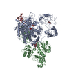
|
|---|---|
| 1 |
|
-Components
-Protein , 2 types, 2 molecules AB
| #1: Protein | Mass: 142162.812 Da / Num. of mol.: 1 / Mutation: N-term truncation and GFP fusion Source method: isolated from a genetically manipulated source Details: Chaetomium thermophilum Endoplasmic reticulum degradation enhancing mannosidase (CtEDEM),Chaetomium thermophilum Endoplasmic reticulum degradation enhancing mannosidase (CtEDEM),Chaetomium ...Details: Chaetomium thermophilum Endoplasmic reticulum degradation enhancing mannosidase (CtEDEM),Chaetomium thermophilum Endoplasmic reticulum degradation enhancing mannosidase (CtEDEM),Chaetomium thermophilum Endoplasmic reticulum degradation enhancing mannosidase (CtEDEM),Chaetomium thermophilum Endoplasmic reticulum degradation enhancing mannosidase (CtEDEM),Chaetomium thermophilum Endoplasmic reticulum degradation enhancing mannosidase (CtEDEM),Chaetomium thermophilum Endoplasmic reticulum degradation enhancing mannosidase (CtEDEM),Chaetomium thermophilum Endoplasmic reticulum degradation enhancing mannosidase (CtEDEM),Chaetomium thermophilum Endoplasmic reticulum degradation enhancing mannosidase (CtEDEM) Source: (gene. exp.) Thermochaetoides thermophila (fungus) / Gene: GFP, CTHT_0058730 / Plasmid: CtEDEM_pHLsec / Cell line (production host): HEK293F / Production host:  Homo sapiens (human) Homo sapiens (human)References: UniProt: P42212, UniProt: G0SCX7, Hydrolases; Glycosylases; Glycosidases, i.e. enzymes that hydrolyse O- and S-glycosyl compounds |
|---|---|
| #2: Protein | Mass: 55841.367 Da / Num. of mol.: 1 / Mutation: Deletion of Cterminal HDEL Source method: isolated from a genetically manipulated source Details: Chaetomium thermophilum Endoplasmic reticulum degradation enhancing protein disulfide isomerase (CtPDI) Source: (gene. exp.) Thermochaetoides thermophila (fungus) / Gene: CTHT_0067360 / Plasmid: CtEDEM_pHLsec / Cell line (production host): HEK293F / Production host: Homo sapiens (human) / References: UniProt: G0SGS2, protein disulfide-isomerase |
-Sugars , 4 types, 6 molecules 

| #3: Polysaccharide | | #4: Polysaccharide | 2-acetamido-2-deoxy-beta-D-glucopyranose-(1-4)-2-acetamido-2-deoxy-beta-D-glucopyranose | Source method: isolated from a genetically manipulated source #7: Sugar |  Type: D-saccharide, beta linking / Mass: 221.208 Da / Num. of mol.: 2 / Source method: obtained synthetically / Formula: C8H15NO6 Type: D-saccharide, beta linking / Mass: 221.208 Da / Num. of mol.: 2 / Source method: obtained synthetically / Formula: C8H15NO6#8: Sugar | ChemComp-MAN / |  Type: D-saccharide, alpha linking / Mass: 180.156 Da / Num. of mol.: 1 / Source method: obtained synthetically / Formula: C6H12O6 Type: D-saccharide, alpha linking / Mass: 180.156 Da / Num. of mol.: 1 / Source method: obtained synthetically / Formula: C6H12O6 |
|---|
-Non-polymers , 3 types, 5 molecules 

| #5: Chemical | ChemComp-CA /  Mass: 40.078 Da / Num. of mol.: 1 Mass: 40.078 Da / Num. of mol.: 1Source method: isolated from a genetically manipulated source Formula: Ca / Feature type: SUBJECT OF INVESTIGATION |
|---|---|
| #6: Chemical | ChemComp-THJ /  Mass: 112.128 Da / Num. of mol.: 1 / Source method: obtained synthetically / Formula: O3S2 Mass: 112.128 Da / Num. of mol.: 1 / Source method: obtained synthetically / Formula: O3S2 |
| #9: Water | ChemComp-HOH / Mass: 18.015 Da / Num. of mol.: 3 / Source method: isolated from a natural source / Formula: H2O |
-Details
| Has ligand of interest | Y |
|---|---|
| Has protein modification | Y |
-Experimental details
-Experiment
| Experiment | Method: ELECTRON MICROSCOPY |
|---|---|
| EM experiment | Aggregation state: PARTICLE / 3D reconstruction method: single particle reconstruction |
- Sample preparation
| Component |
| ||||||||||||||||||||||||||||
|---|---|---|---|---|---|---|---|---|---|---|---|---|---|---|---|---|---|---|---|---|---|---|---|---|---|---|---|---|---|
| Molecular weight |
| ||||||||||||||||||||||||||||
| Source (natural) |
| ||||||||||||||||||||||||||||
| Source (recombinant) |
| ||||||||||||||||||||||||||||
| Buffer solution | pH: 7 Details: Micro SEC buffer: 150 mM NaCl, 20 mM MES pH 7.0, 1 mM CaCl2 | ||||||||||||||||||||||||||||
| Buffer component |
| ||||||||||||||||||||||||||||
| Specimen | Conc.: 0.02 mg/ml / Embedding applied: NO / Shadowing applied: NO / Staining applied: NO / Vitrification applied: YES Details: 40 uL of a protein sample with an OD_280 of 0.88 were injected onto a Cytiva Superdex 200 Increase 3.2/300 size-exclusion chromatography column, equilibrated in 150 mM NaCl, 20 mM MES pH 7. ...Details: 40 uL of a protein sample with an OD_280 of 0.88 were injected onto a Cytiva Superdex 200 Increase 3.2/300 size-exclusion chromatography column, equilibrated in 150 mM NaCl, 20 mM MES pH 7.0, 1 mM CaCl2, collecting 50 uL fractions. | ||||||||||||||||||||||||||||
| Specimen support | Details: The Quantifoil Au300 R1.2/1.3 Holey Carbon grids were glow discharged using the EMS GloQube. For grids to be coated in GO, the Graphene Oxide setting was used (0.2m Bar, 40 mA for 180 ...Details: The Quantifoil Au300 R1.2/1.3 Holey Carbon grids were glow discharged using the EMS GloQube. For grids to be coated in GO, the Graphene Oxide setting was used (0.2m Bar, 40 mA for 180 seconds). For GO coating, graphene oxide solution (Sigma) at 0.2 mg/ml was spun at 300 g for 30 s to remove large aggregates. 3 uL was added to the grids for 1 minute, before blotting with Whatman No1 filter paper and washing three times with 20 uL drops of ultrapure water. Grid material: GOLD / Grid mesh size: 300 divisions/in. / Grid type: Quantifoil R1.2/1.3 | ||||||||||||||||||||||||||||
| Vitrification | Instrument: FEI VITROBOT MARK IV / Cryogen name: ETHANE / Humidity: 100 % / Chamber temperature: 277 K Details: Blotting time 3 s, waiting time 30 s Sample volume 3 uL, blot force 10 |
- Electron microscopy imaging
| Experimental equipment |  Model: Titan Krios / Image courtesy: FEI Company |
|---|---|
| Microscopy | Model: FEI TITAN KRIOS |
| Electron gun | Electron source:  FIELD EMISSION GUN / Accelerating voltage: 300 kV / Illumination mode: FLOOD BEAM FIELD EMISSION GUN / Accelerating voltage: 300 kV / Illumination mode: FLOOD BEAM |
| Electron lens | Mode: BRIGHT FIELD / Nominal magnification: 105000 X / Nominal defocus max: 2500 nm / Nominal defocus min: 1000 nm / Cs: 2.7 mm / C2 aperture diameter: 50 µm / Alignment procedure: BASIC |
| Specimen holder | Cryogen: NITROGEN / Specimen holder model: FEI TITAN KRIOS AUTOGRID HOLDER |
| Image recording | Electron dose: 1 e/Å2 / Film or detector model: GATAN K3 (6k x 4k) |
- Processing
| EM software |
| ||||||||||||||||||||||||
|---|---|---|---|---|---|---|---|---|---|---|---|---|---|---|---|---|---|---|---|---|---|---|---|---|---|
| CTF correction | Type: PHASE FLIPPING AND AMPLITUDE CORRECTION | ||||||||||||||||||||||||
| Symmetry | Point symmetry: C1 (asymmetric) | ||||||||||||||||||||||||
| 3D reconstruction | Resolution: 2.6 Å / Resolution method: FSC 0.143 CUT-OFF / Num. of particles: 1400000 / Algorithm: BACK PROJECTION / Num. of class averages: 4 / Symmetry type: POINT | ||||||||||||||||||||||||
| Refine LS restraints |
|
