 Movie
Movie Controller

[English] 日本語
 Yorodumi
- PDB-8pi5: Crystal structure of human insulin desB30 precursor with an Alani... -
+ Open data

- Basic information
| Entry | Database: PDB / ID: 8pi5 | ||||||
|---|---|---|---|---|---|---|---|
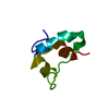
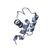
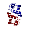
| Title | Crystal structure of human insulin desB30 precursor with an Alanine-Methionine-Lysine C-peptide in hexamer (T3R3) conformation | ||||||
 Components Components | Insulin B chain,Insulin A chain | ||||||
 Keywords Keywords | HORMONE / insulin / precursor / dimer | ||||||
| Function / homology |  Function and homology information Function and homology information: / negative regulation of glycogen catabolic process / negative regulation of fatty acid metabolic process / Signaling by Insulin receptor / IRS activation / Insulin processing / regulation of protein secretion / positive regulation of peptide hormone secretion / negative regulation of feeding behavior / negative regulation of acute inflammatory response ...: / negative regulation of glycogen catabolic process / negative regulation of fatty acid metabolic process / Signaling by Insulin receptor / IRS activation / Insulin processing / regulation of protein secretion / positive regulation of peptide hormone secretion / negative regulation of feeding behavior / negative regulation of acute inflammatory response / Regulation of gene expression in beta cells / positive regulation of respiratory burst / alpha-beta T cell activation / Synthesis, secretion, and deacylation of Ghrelin / negative regulation of protein secretion / negative regulation of gluconeogenesis / positive regulation of dendritic spine maintenance / fatty acid homeostasis / positive regulation of glycogen biosynthetic process / positive regulation of insulin receptor signaling pathway / Signal attenuation / FOXO-mediated transcription of oxidative stress, metabolic and neuronal genes / positive regulation of lipid biosynthetic process / negative regulation of respiratory burst involved in inflammatory response / negative regulation of lipid catabolic process / negative regulation of oxidative stress-induced intrinsic apoptotic signaling pathway / nitric oxide-cGMP-mediated signaling / regulation of protein localization to plasma membrane / transport vesicle / Insulin receptor recycling / COPI-mediated anterograde transport / positive regulation of nitric-oxide synthase activity / negative regulation of reactive oxygen species biosynthetic process / positive regulation of brown fat cell differentiation / insulin-like growth factor receptor binding / NPAS4 regulates expression of target genes / neuron projection maintenance / positive regulation of mitotic nuclear division / endoplasmic reticulum-Golgi intermediate compartment membrane / positive regulation of glycolytic process / Insulin receptor signalling cascade / positive regulation of D-glucose import across plasma membrane / endosome lumen / positive regulation of protein secretion / acute-phase response / positive regulation of cytokine production / insulin receptor binding / wound healing / positive regulation of long-term synaptic potentiation / positive regulation of cell differentiation / positive regulation of neuron projection development / negative regulation of protein catabolic process / Regulation of insulin secretion / hormone activity / positive regulation of protein localization to nucleus / glucose metabolic process / regulation of synaptic plasticity / vasodilation / Golgi lumen / cognition / glucose homeostasis / insulin receptor signaling pathway / regulation of protein localization / cell-cell signaling / PI5P, PP2A and IER3 Regulate PI3K/AKT Signaling / positive regulation of cell growth / protease binding / secretory granule lumen / positive regulation of MAPK cascade / positive regulation of phosphatidylinositol 3-kinase/protein kinase B signal transduction / positive regulation of canonical NF-kappaB signal transduction / positive regulation of cell migration / G protein-coupled receptor signaling pathway / endoplasmic reticulum lumen / Amyloid fiber formation / receptor ligand activity / negative regulation of gene expression / Golgi membrane / positive regulation of gene expression / positive regulation of cell population proliferation / regulation of DNA-templated transcription / : / extracellular region / identical protein binding Similarity search - Function | ||||||
| Biological species |  Homo sapiens (human) Homo sapiens (human) | ||||||
| Method |  X-RAY DIFFRACTION / MOLECULAR REPLACEMENT / Resolution: 1.66 Å X-RAY DIFFRACTION / MOLECULAR REPLACEMENT / Resolution: 1.66 Å | ||||||
 Authors Authors | Johansson, E. / Schluckebier, G. | ||||||
| Funding support |  Denmark, 1items Denmark, 1items
| ||||||
 Citation Citation | Journal: Trends Biotechnol / Year: 2024 Title: Molecular engineering of insulin for recombinant expression in yeast. Authors: Kjeldsen, T. / Andersen, A.S. / Hubalek, F. / Johansson, E. / Kreiner, F.F. / Schluckebier, G. / Kurtzhals, P. | ||||||
| History |
|
- Structure visualization
| Structure viewer | Molecule: MolmilJmol/JSmol |
|---|
- Downloads & links
-Download
| PDBx/mmCIF format | 8pi5.cif.gz | 81.4 KB | Display | PDBx/mmCIF format |
|---|---|---|---|---|
| PDB format | pdb8pi5.ent.gz | 58.7 KB | Display | PDB format |
| PDBx/mmJSON format | 8pi5.json.gz | Tree view | PDBx/mmJSON format | |
| Others |  Other downloads Other downloads |
-Validation report
| Arichive directory | https://data.pdbj.org/pub/pdb/validation_reports/pi/8pi5ftp://data.pdbj.org/pub/pdb/validation_reports/pi/8pi5 | HTTPS FTP |
|---|
-Related structure data

-Links
 PDBj
PDBj


- Assembly
| Deposited unit | 
| |||||||||||||||||||||||||||
|---|---|---|---|---|---|---|---|---|---|---|---|---|---|---|---|---|---|---|---|---|---|---|---|---|---|---|---|---|
| 1 | 
| |||||||||||||||||||||||||||
| Unit cell |
| |||||||||||||||||||||||||||
| Components on special symmetry positions |
|
-Components
| #1: Protein | Mass: 6029.985 Da / Num. of mol.: 2 Source method: isolated from a genetically manipulated source Source: (gene. exp.) Homo sapiens (human) / Gene: INS / Production host:  #2: Chemical | ChemComp-ZN /   Mass: 65.409 Da / Num. of mol.: 4 / Source method: obtained synthetically / Formula: Zn Mass: 65.409 Da / Num. of mol.: 4 / Source method: obtained synthetically / Formula: Zn#3: Chemical | ChemComp-CL / |   Mass: 35.453 Da / Num. of mol.: 1 / Source method: obtained synthetically / Formula: Cl Mass: 35.453 Da / Num. of mol.: 1 / Source method: obtained synthetically / Formula: Cl#4: Chemical | ChemComp-RCO / |   Mass: 110.111 Da / Num. of mol.: 1 / Source method: obtained synthetically / Formula: C6H6O2 Mass: 110.111 Da / Num. of mol.: 1 / Source method: obtained synthetically / Formula: C6H6O2#5: Water | ChemComp-HOH / |  Mass: 18.015 Da / Num. of mol.: 94 / Source method: isolated from a natural source / Formula: H2O Mass: 18.015 Da / Num. of mol.: 94 / Source method: isolated from a natural source / Formula: H2OHas ligand of interest | N | Has protein modification | Y | |
|---|
-Experimental details
-Experiment
| Experiment | Method: X-RAY DIFFRACTION / Number of used crystals: 1 |
|---|
- Sample preparation
| Crystal | Density Matthews: 1.97 Å3/Da / Density % sol: 37.52 % |
|---|---|
| Crystal grow | Temperature: 291 K / Method: vapor diffusion, sitting drop / pH: 9 Details: 6 mg/ml protein, 20 mM resorcinol, 0.5 Zn2+ (from zinc acetate) per insuln monomer in water, pH 7.95 precipitant: 0.1 M Bicine, pH 9.0, 2 % (v/v) 1,4-dioxane, 10 % (w/v) PEG 20000 |
-Data collection
| Diffraction | Mean temperature: 100 K / Serial crystal experiment: N |
|---|---|
| Diffraction source | Source: ROTATING ANODE / Type: RIGAKU MICROMAX-007 HF / Wavelength: 1.542 Å |
| Detector | Type: MAR scanner 345 mm plate / Detector: IMAGE PLATE / Date: Jan 22, 2013 |
| Radiation | Protocol: SINGLE WAVELENGTH / Monochromatic (M) / Laue (L): M / Scattering type: x-ray |
| Radiation wavelength | Wavelength: 1.542 Å / Relative weight: 1 |
| Reflection | Resolution: 1.66→13.66 Å / Num. obs: 10563 / % possible obs: 97.17 % / Redundancy: 6.1 % / Biso Wilson estimate: 21.26 Å2 / CC1/2: 0.999 / CC star: 1 / Rmerge(I) obs: 0.04731 / Rpim(I) all: 0.01975 / Rrim(I) all: 0.05135 / Net I/σ(I): 25.54 |
| Reflection shell | Resolution: 1.66→1.72 Å / Redundancy: 2.5 % / Rmerge(I) obs: 0.3121 / Mean I/σ(I) obs: 3.58 / Num. unique obs: 829 / CC1/2: 0.847 / CC star: 0.958 / Rpim(I) all: 0.2031 / Rrim(I) all: 0.375 / % possible all: 76.19 |
- Processing
| Software |
| ||||||||||||||||||||||||||||||||||||||||||||||||||||||||
|---|---|---|---|---|---|---|---|---|---|---|---|---|---|---|---|---|---|---|---|---|---|---|---|---|---|---|---|---|---|---|---|---|---|---|---|---|---|---|---|---|---|---|---|---|---|---|---|---|---|---|---|---|---|---|---|---|---|
| Refinement | Method to determine structure: MOLECULAR REPLACEMENT / Resolution: 1.66→13.66 Å / SU ML: 0.1539 / Cross valid method: FREE R-VALUE / σ(F): 1.96 / Phase error: 22.8241 Stereochemistry target values: GeoStd + Monomer Library + CDL v1.2
| ||||||||||||||||||||||||||||||||||||||||||||||||||||||||
| Solvent computation | Shrinkage radii: 0.9 Å / VDW probe radii: 1.11 Å / Solvent model: FLAT BULK SOLVENT MODEL | ||||||||||||||||||||||||||||||||||||||||||||||||||||||||
| Displacement parameters | Biso mean: 29.36 Å2 | ||||||||||||||||||||||||||||||||||||||||||||||||||||||||
| Refinement step | Cycle: LAST / Resolution: 1.66→13.66 Å
| ||||||||||||||||||||||||||||||||||||||||||||||||||||||||
| Refine LS restraints |
| ||||||||||||||||||||||||||||||||||||||||||||||||||||||||
| LS refinement shell |
|
