 Movie
Movie Controller

[English] 日本語
 Yorodumi
- PDB-8pea: OXA-48_F72L. Epistasis Arises from Shifting the Rate-Limiting Ste... -
+ Open data

- Basic information
| Entry | Database: PDB / ID: 8pea | ||||||
|---|---|---|---|---|---|---|---|
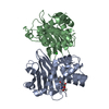
| Title | OXA-48_F72L. Epistasis Arises from Shifting the Rate-Limiting Step during Enzyme Evolution | ||||||
 Components Components | Beta-lactamase | ||||||
 Keywords Keywords | HYDROLASE / Protein evolution. Antibiotic resistance. OXA-48. | ||||||
| Function / homology |  Function and homology information Function and homology informationpenicillin binding / antibiotic catabolic process / cell wall organization / beta-lactamase activity / beta-lactamase / response to antibiotic / plasma membrane Similarity search - Function | ||||||
| Biological species |  Klebsiella pneumoniae (bacteria) Klebsiella pneumoniae (bacteria) | ||||||
| Method |  X-RAY DIFFRACTION / SYNCHROTRON / MOLECULAR REPLACEMENT / Resolution: 1.97 Å X-RAY DIFFRACTION / SYNCHROTRON / MOLECULAR REPLACEMENT / Resolution: 1.97 Å | ||||||
 Authors Authors | Leiros, H.-K.S. / Frohlich, C. | ||||||
| Funding support | 1items
| ||||||
 Citation Citation | Journal: Nat Catal / Year: 2024 Title: Epistasis arises from shifting the rate-limiting step during enzyme evolution of a beta-lactamase. Authors: Frohlich, C. / Bunzel, H.A. / Buda, K. / Mulholland, A.J. / van der Kamp, M.W. / Johnsen, P.J. / Leiros, H.S. / Tokuriki, N. | ||||||
| History |
|
- Structure visualization
| Structure viewer | Molecule: MolmilJmol/JSmol |
|---|
- Downloads & links
-Download
| PDBx/mmCIF format | 8pea.cif.gz | 120.8 KB | Display | PDBx/mmCIF format |
|---|---|---|---|---|
| PDB format | pdb8pea.ent.gz | 90.6 KB | Display | PDB format |
| PDBx/mmJSON format | 8pea.json.gz | Tree view | PDBx/mmJSON format | |
| Others |  Other downloads Other downloads |
-Validation report
| Arichive directory | https://data.pdbj.org/pub/pdb/validation_reports/pe/8peaftp://data.pdbj.org/pub/pdb/validation_reports/pe/8pea | HTTPS FTP |
|---|
-Related structure data

-Links
 PDBj
PDBj- Assembly
| Deposited unit | 
| ||||||||||||
|---|---|---|---|---|---|---|---|---|---|---|---|---|---|
| 1 |
| ||||||||||||
| Unit cell |
|
-Components
| #1: Protein | Mass: 30362.709 Da / Num. of mol.: 2 Source method: isolated from a genetically manipulated source Details: This is a F72L mutant / Source: (gene. exp.) Klebsiella pneumoniae (bacteria) / Gene: bla OXA-48 / Production host: #2: Chemical | ChemComp-CL / |   Mass: 35.453 Da / Num. of mol.: 1 / Source method: obtained synthetically / Formula: Cl Mass: 35.453 Da / Num. of mol.: 1 / Source method: obtained synthetically / Formula: Cl#3: Water | ChemComp-HOH / |  Mass: 18.015 Da / Num. of mol.: 295 / Source method: isolated from a natural source / Formula: H2O Mass: 18.015 Da / Num. of mol.: 295 / Source method: isolated from a natural source / Formula: H2OHas ligand of interest | N | |
|---|
-Experimental details
-Experiment
| Experiment | Method: X-RAY DIFFRACTION / Number of used crystals: 1 |
|---|
- Sample preparation
| Crystal | Density Matthews: 2.23 Å3/Da / Density % sol: 44.77 % |
|---|---|
| Crystal grow | Temperature: 277 K / Method: vapor diffusion, hanging drop / pH: 9 Details: 0.1 M Tris, pH 9.0 28-30% polyethylene glycol (PEG) mono ethylene ether 500 277 K. 10 mg/mL protein |
-Data collection
| Diffraction | Mean temperature: 100 K / Serial crystal experiment: N |
|---|---|
| Diffraction source | Source: SYNCHROTRON / Site: ESRF  / Beamline: ID23-2 / Wavelength: 0.873128 Å / Beamline: ID23-2 / Wavelength: 0.873128 Å |
| Detector | Type: DECTRIS PILATUS 6M / Detector: PIXEL / Date: Apr 18, 2021 |
| Radiation | Protocol: SINGLE WAVELENGTH / Monochromatic (M) / Laue (L): M / Scattering type: x-ray |
| Radiation wavelength | Wavelength: 0.873128 Å / Relative weight: 1 |
| Reflection | Resolution: 1.97→44.41 Å / Num. obs: 38991 / % possible obs: 99.75 % / Redundancy: 7.1 % / Biso Wilson estimate: 37.28 Å2 / CC1/2: 0.998 / Rmerge(I) obs: 0.1072 / Rpim(I) all: 0.04355 / Rrim(I) all: 0.1159 / Net I/σ(I): 19.05 |
| Reflection shell | Resolution: 1.97→2.04 Å / Redundancy: 6.5 % / Rmerge(I) obs: 1.352 / Mean I/σ(I) obs: 0.99 / Num. unique obs: 3822 / CC1/2: 0.898 / Rpim(I) all: 0.5769 / Rrim(I) all: 1.474 / % possible all: 99.74 |
- Processing
| Software |
| |||||||||||||||||||||||||||||||||||||||||||||||||||||||||||||||||||||||||||||||||||||||||||||||||||||||||||||||||||||||||||||||||||||||||||||||||||||||||||||||||
|---|---|---|---|---|---|---|---|---|---|---|---|---|---|---|---|---|---|---|---|---|---|---|---|---|---|---|---|---|---|---|---|---|---|---|---|---|---|---|---|---|---|---|---|---|---|---|---|---|---|---|---|---|---|---|---|---|---|---|---|---|---|---|---|---|---|---|---|---|---|---|---|---|---|---|---|---|---|---|---|---|---|---|---|---|---|---|---|---|---|---|---|---|---|---|---|---|---|---|---|---|---|---|---|---|---|---|---|---|---|---|---|---|---|---|---|---|---|---|---|---|---|---|---|---|---|---|---|---|---|---|---|---|---|---|---|---|---|---|---|---|---|---|---|---|---|---|---|---|---|---|---|---|---|---|---|---|---|---|---|---|---|---|
| Refinement | Method to determine structure: MOLECULAR REPLACEMENT / Resolution: 1.97→25 Å / SU ML: 0.3087 / Cross valid method: FREE R-VALUE / σ(F): 1.34 / Phase error: 30.4596 Stereochemistry target values: GeoStd + Monomer Library + CDL v1.2
| |||||||||||||||||||||||||||||||||||||||||||||||||||||||||||||||||||||||||||||||||||||||||||||||||||||||||||||||||||||||||||||||||||||||||||||||||||||||||||||||||
| Solvent computation | Shrinkage radii: 0.9 Å / VDW probe radii: 1.11 Å / Solvent model: FLAT BULK SOLVENT MODEL | |||||||||||||||||||||||||||||||||||||||||||||||||||||||||||||||||||||||||||||||||||||||||||||||||||||||||||||||||||||||||||||||||||||||||||||||||||||||||||||||||
| Displacement parameters | Biso mean: 45.74 Å2 | |||||||||||||||||||||||||||||||||||||||||||||||||||||||||||||||||||||||||||||||||||||||||||||||||||||||||||||||||||||||||||||||||||||||||||||||||||||||||||||||||
| Refinement step | Cycle: LAST / Resolution: 1.97→25 Å
| |||||||||||||||||||||||||||||||||||||||||||||||||||||||||||||||||||||||||||||||||||||||||||||||||||||||||||||||||||||||||||||||||||||||||||||||||||||||||||||||||
| Refine LS restraints |
| |||||||||||||||||||||||||||||||||||||||||||||||||||||||||||||||||||||||||||||||||||||||||||||||||||||||||||||||||||||||||||||||||||||||||||||||||||||||||||||||||
| LS refinement shell |
|
