 Movie
Movie Controller

[English] 日本語
 Yorodumi
- PDB-8pdr: Rigid body fit of assembled HMPV N-RNA spiral bound to the C-term... -
+ Open data

- Basic information
| Entry | Database: PDB / ID: 8pdr | |||||||||||||||||||||||||||||||||||||||
|---|---|---|---|---|---|---|---|---|---|---|---|---|---|---|---|---|---|---|---|---|---|---|---|---|---|---|---|---|---|---|---|---|---|---|---|---|---|---|---|---|
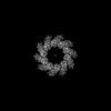
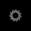
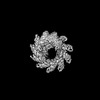
| Title | Rigid body fit of assembled HMPV N-RNA spiral bound to the C-terminal region of P | |||||||||||||||||||||||||||||||||||||||
 Components Components |
| |||||||||||||||||||||||||||||||||||||||
 Keywords Keywords | VIRAL PROTEIN / Nucleoprotein / Virus / Nucleocapsid / RNA-binding / HMPV / Pneumoviridae / Mononegavirales | |||||||||||||||||||||||||||||||||||||||
| Function / homology |  Function and homology information Function and homology informationhelical viral capsid / virion component / viral nucleocapsid / host cell cytoplasm / ribonucleoprotein complex / RNA-directed RNA polymerase activity / RNA binding Similarity search - Function | |||||||||||||||||||||||||||||||||||||||
| Biological species |  Human metapneumovirus Human metapneumovirus | |||||||||||||||||||||||||||||||||||||||
| Method | ELECTRON MICROSCOPY / single particle reconstruction / cryo EM / Resolution: 4 Å | |||||||||||||||||||||||||||||||||||||||
 Authors Authors | Whitehead, J.D. / Decool, H. / Leyrat, C. / Carrique, L. / Fix, J. / Eleouet, J.F. / Galloux, M. / Renner, M. | |||||||||||||||||||||||||||||||||||||||
| Funding support |  United Kingdom, 1items United Kingdom, 1items
| |||||||||||||||||||||||||||||||||||||||
 Citation Citation | Journal: Nat Commun / Year: 2023 Title: Structure of the N-RNA/P interface indicates mode of L/P recruitment to the nucleocapsid of human metapneumovirus. Authors: Jack D Whitehead / Hortense Decool / Cédric Leyrat / Loic Carrique / Jenna Fix / Jean-François Eléouët / Marie Galloux / Max Renner /   Abstract: Human metapneumovirus (HMPV) is a major cause of respiratory illness in young children. The HMPV polymerase (L) binds an obligate cofactor, the phosphoprotein (P). During replication and ...Human metapneumovirus (HMPV) is a major cause of respiratory illness in young children. The HMPV polymerase (L) binds an obligate cofactor, the phosphoprotein (P). During replication and transcription, the L/P complex traverses the viral RNA genome, which is encapsidated within nucleoproteins (N). An essential interaction between N and a C-terminal region of P tethers the L/P polymerase to the template. This N-P interaction is also involved in the formation of cytoplasmic viral factories in infected cells, called inclusion bodies. To define how the polymerase component P recognizes N-encapsidated RNA (N-RNA) we employed cryogenic electron microscopy (cryo-EM) and molecular dynamics simulations, coupled to activity assays and imaging of inclusion bodies in cells. We report a 2.9 Å resolution structure of a triple-complex between multimeric N, bound to both RNA and the C-terminal region of P. Furthermore, we also present cryo-EM structures of assembled N in different oligomeric states, highlighting the plasticity of N. Combined with our functional assays, these structural data delineate in molecular detail how P attaches to N-RNA whilst retaining substantial conformational dynamics. Moreover, the N-RNA-P triple complex structure provides a molecular blueprint for the design of therapeutics to potentially disrupt the attachment of L/P to its template. | |||||||||||||||||||||||||||||||||||||||
| History |
|
- Structure visualization
| Structure viewer | Molecule: MolmilJmol/JSmol |
|---|
- Downloads & links
-Download
| PDBx/mmCIF format | 8pdr.cif.gz | 676.9 KB | Display | PDBx/mmCIF format |
|---|---|---|---|---|
| PDB format | pdb8pdr.ent.gz | 571.4 KB | Display | PDB format |
| PDBx/mmJSON format | 8pdr.json.gz | Tree view | PDBx/mmJSON format | |
| Others |  Other downloads Other downloads |
-Validation report
| Arichive directory | https://data.pdbj.org/pub/pdb/validation_reports/pd/8pdrftp://data.pdbj.org/pub/pdb/validation_reports/pd/8pdr | HTTPS FTP |
|---|
-Related structure data
| Related structure data |  17619MC  8pdlC  8pdmC  8pdnC  8pdoC  8pdpC  8pdqC  8pdsC M: map data used to model this data C: citing same article ( |
|---|---|
| Similar structure data |


-Links
 PDBj
PDBj- Assembly
| Deposited unit | 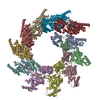
|
|---|---|
| 1 |
|
-Components
| #1: Protein | Mass: 44534.570 Da / Num. of mol.: 11 Source method: isolated from a genetically manipulated source Source: (gene. exp.) Human metapneumovirus (strain CAN97-83)Strain: CAN97-83 / Gene: N / Production host: #2: Protein/peptide | Mass: 1139.276 Da / Num. of mol.: 11 / Source method: obtained synthetically / Source: (synth.) Human metapneumovirus (strain CAN97-83) / References: UniProt: Q8B9Q8#3: RNA chain | Mass: 2091.315 Da / Num. of mol.: 11 / Source method: isolated from a natural source / Source: (natural) Has protein modification | N | |
|---|
-Experimental details
-Experiment
| Experiment | Method: ELECTRON MICROSCOPY |
|---|---|
| EM experiment | Aggregation state: PARTICLE / 3D reconstruction method: single particle reconstruction |
- Sample preparation
| Component |
| ||||||||||||||||||||||||||||||
|---|---|---|---|---|---|---|---|---|---|---|---|---|---|---|---|---|---|---|---|---|---|---|---|---|---|---|---|---|---|---|---|
| Molecular weight | Experimental value: NO | ||||||||||||||||||||||||||||||
| Source (natural) |
| ||||||||||||||||||||||||||||||
| Source (recombinant) |
| ||||||||||||||||||||||||||||||
| Buffer solution | pH: 8 / Details: 12.5 mM Tris-HCl, pH 8.0, 125 mM NaCl | ||||||||||||||||||||||||||||||
| Specimen | Conc.: 0.5 mg/ml / Embedding applied: NO / Shadowing applied: NO / Staining applied: NO / Vitrification applied: YES | ||||||||||||||||||||||||||||||
| Specimen support | Grid material: COPPER / Grid type: Quantifoil R2/1 | ||||||||||||||||||||||||||||||
| Vitrification | Instrument: FEI VITROBOT MARK IV / Cryogen name: ETHANE-PROPANE |
- Electron microscopy imaging
| Experimental equipment |  Model: Titan Krios / Image courtesy: FEI Company |
|---|---|
| Microscopy | Model: TFS KRIOS |
| Electron gun | Electron source:  FIELD EMISSION GUN / Accelerating voltage: 300 kV / Illumination mode: FLOOD BEAM FIELD EMISSION GUN / Accelerating voltage: 300 kV / Illumination mode: FLOOD BEAM |
| Electron lens | Mode: BRIGHT FIELD / Nominal defocus max: 2600 nm / Nominal defocus min: 1000 nm |
| Specimen holder | Specimen holder model: FEI TITAN KRIOS AUTOGRID HOLDER |
| Image recording | Electron dose: 44.2 e/Å2 / Film or detector model: GATAN K2 SUMMIT (4k x 4k) |
- Processing
| EM software |
| ||||||||||||||||||||||||
|---|---|---|---|---|---|---|---|---|---|---|---|---|---|---|---|---|---|---|---|---|---|---|---|---|---|
| CTF correction | Type: PHASE FLIPPING AND AMPLITUDE CORRECTION | ||||||||||||||||||||||||
| Particle selection | Details: Please see publication for details | ||||||||||||||||||||||||
| Symmetry | Point symmetry: C1 (asymmetric) | ||||||||||||||||||||||||
| 3D reconstruction | Resolution: 4 Å / Resolution method: FSC 0.143 CUT-OFF / Num. of particles: 201568 / Symmetry type: POINT | ||||||||||||||||||||||||
| Atomic model building | Protocol: RIGID BODY FIT / Details: Rigid body only | ||||||||||||||||||||||||
| Refine LS restraints |
|
