 Movie
Movie Controller

[English] 日本語
 Yorodumi
- PDB-8pcm: Structure of serine-beta-lactamase CTX-M-14 following the time-re... -
+ Open data

- Basic information
| Entry | Database: PDB / ID: 8pcm | ||||||||||||
|---|---|---|---|---|---|---|---|---|---|---|---|---|---|
| Title | Structure of serine-beta-lactamase CTX-M-14 following the time-resolved active site binding of boric acid and subsequent glycerol-boric acid-ester formation, 80 ms | ||||||||||||
 Components Components | Beta-lactamase | ||||||||||||
 Keywords Keywords | HYDROLASE / catalytic activity / beta-lactamase activity / hydrolase activity / inhibitor / time-resolved / tape drive | ||||||||||||
| Function / homology |  Function and homology information Function and homology informationbeta-lactam antibiotic catabolic process / beta-lactamase activity / beta-lactamase / response to antibiotic Similarity search - Function | ||||||||||||
| Biological species |  Klebsiella pneumoniae (bacteria) Klebsiella pneumoniae (bacteria) | ||||||||||||
| Method |  X-RAY DIFFRACTION / SYNCHROTRON / MOLECULAR REPLACEMENT / Resolution: 1.84 Å X-RAY DIFFRACTION / SYNCHROTRON / MOLECULAR REPLACEMENT / Resolution: 1.84 Å | ||||||||||||
 Authors Authors | Prester, A. / Perbandt, M. / Galchenkova, M. / Oberthuer, D. / Yefanov, O. / Hinrichs, W. / Rohde, H. / Betzel, C. | ||||||||||||
| Funding support |  Germany, 3items Germany, 3items
| ||||||||||||
 Citation Citation | Journal: Commun Chem / Year: 2024 Title: Time-resolved crystallography of boric acid binding to the active site serine of the beta-lactamase CTX-M-14 and subsequent 1,2-diol esterification. Authors: Prester, A. / Perbandt, M. / Galchenkova, M. / Oberthuer, D. / Werner, N. / Henkel, A. / Maracke, J. / Yefanov, O. / Hakanpaa, J. / Pompidor, G. / Meyer, J. / Chapman, H. / Aepfelbacher, M. ...Authors: Prester, A. / Perbandt, M. / Galchenkova, M. / Oberthuer, D. / Werner, N. / Henkel, A. / Maracke, J. / Yefanov, O. / Hakanpaa, J. / Pompidor, G. / Meyer, J. / Chapman, H. / Aepfelbacher, M. / Hinrichs, W. / Rohde, H. / Betzel, C. | ||||||||||||
| History |
|
- Structure visualization
| Structure viewer | Molecule: MolmilJmol/JSmol |
|---|
- Downloads & links
-Download
| PDBx/mmCIF format | 8pcm.cif.gz | 172.8 KB | Display | PDBx/mmCIF format |
|---|---|---|---|---|
| PDB format | pdb8pcm.ent.gz | 131.6 KB | Display | PDB format |
| PDBx/mmJSON format | 8pcm.json.gz | Tree view | PDBx/mmJSON format | |
| Others |  Other downloads Other downloads |
-Validation report
| Arichive directory | https://data.pdbj.org/pub/pdb/validation_reports/pc/8pcmftp://data.pdbj.org/pub/pdb/validation_reports/pc/8pcm | HTTPS FTP |
|---|
-Related structure data
| Related structure data | 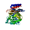 8pc9C  8pcaC 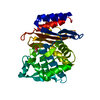 8pcbC  8pccC  8pcdC 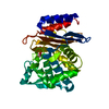 8pceC  8pcfC  8pcgC  8pciC  8pcjC  8pckC  8pclC 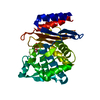 8pcnC  8pcoC  8pcpC  8pcqC  8pcrC  8pcsC  8pctC  8pcuC  8pcvC 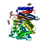 8r7mC C: citing same article ( |
|---|---|
| Similar structure data |
-Links
 PDBj
PDBj


- Assembly
| Deposited unit | 
| ||||||||||||
|---|---|---|---|---|---|---|---|---|---|---|---|---|---|
| 1 |
| ||||||||||||
| Unit cell |
|
-Components
| #1: Protein | Mass: 28000.547 Da / Num. of mol.: 1 Source method: isolated from a genetically manipulated source Source: (gene. exp.) Klebsiella pneumoniae (bacteria) / Gene: ctx-m-14 / Production host: |
|---|---|
| #2: Chemical | ChemComp-YCH / [(  Mass: 117.896 Da / Num. of mol.: 1 / Source method: obtained synthetically / Formula: C3H7BO4 / Feature type: SUBJECT OF INVESTIGATION Mass: 117.896 Da / Num. of mol.: 1 / Source method: obtained synthetically / Formula: C3H7BO4 / Feature type: SUBJECT OF INVESTIGATION |
| #3: Chemical | ChemComp-BO4 /   Mass: 78.840 Da / Num. of mol.: 1 / Source method: obtained synthetically / Formula: BH4O4 / Feature type: SUBJECT OF INVESTIGATION Mass: 78.840 Da / Num. of mol.: 1 / Source method: obtained synthetically / Formula: BH4O4 / Feature type: SUBJECT OF INVESTIGATION |
| #4: Chemical | ChemComp-SO4 /   Mass: 96.063 Da / Num. of mol.: 1 / Source method: obtained synthetically / Formula: SO4 Mass: 96.063 Da / Num. of mol.: 1 / Source method: obtained synthetically / Formula: SO4 |
| #5: Water | ChemComp-HOH /  Mass: 18.015 Da / Num. of mol.: 209 / Source method: isolated from a natural source / Formula: H2O Mass: 18.015 Da / Num. of mol.: 209 / Source method: isolated from a natural source / Formula: H2O |
| Has ligand of interest | Y |
| Has protein modification | Y |
-Experimental details
-Experiment
| Experiment | Method: X-RAY DIFFRACTION / Number of used crystals: 1 |
|---|
- Sample preparation
| Crystal | Density Matthews: 2.11 Å3/Da / Density % sol: 41.57 % |
|---|---|
| Crystal grow | Temperature: 293 K / Method: batch mode Details: 50% CTX-M-14 solution (22 mg/ml) was mixed with 45% precipitant solution (40% PEG8000, 200mM lithium sulfate, 100mM sodium acetate, pH 4.5) and with 5% undiluted seed stock in batch crystallization setups |
-Data collection
| Diffraction | Mean temperature: 295 K / Serial crystal experiment: Y |
|---|---|
| Diffraction source | Source: SYNCHROTRON / Site: PETRA III, DESY / Beamline: P11 / Wavelength: 1.033 Å |
| Detector | Type: DECTRIS EIGER2 X 16M / Detector: PIXEL / Date: Mar 27, 2021 |
| Radiation | Protocol: SINGLE WAVELENGTH / Monochromatic (M) / Laue (L): M / Scattering type: x-ray |
| Radiation wavelength | Wavelength: 1.033 Å / Relative weight: 1 |
| Reflection | Resolution: 1.84→38.88 Å / Num. obs: 21824 / % possible obs: 100 % / Redundancy: 3113 % / Biso Wilson estimate: 38.76 Å2 / CC1/2: 0.998 / CC star: 0.999 / Net I/σ(I): 10.73 |
| Reflection shell | Resolution: 1.84→1.86 Å / Num. unique obs: 1402 / CC1/2: 0.195 / CC star: 0.572 |
| Serial crystallography sample delivery | Description: CFEL TapeDrive / Method: injection |
- Processing
| Software |
| ||||||||||||||||||||||||||||||||||||||||||||||||||||||||||||||||||||||
|---|---|---|---|---|---|---|---|---|---|---|---|---|---|---|---|---|---|---|---|---|---|---|---|---|---|---|---|---|---|---|---|---|---|---|---|---|---|---|---|---|---|---|---|---|---|---|---|---|---|---|---|---|---|---|---|---|---|---|---|---|---|---|---|---|---|---|---|---|---|---|---|
| Refinement | Method to determine structure: MOLECULAR REPLACEMENT / Resolution: 1.84→38.88 Å / SU ML: 0.3215 / Cross valid method: FREE R-VALUE / σ(F): 1.34 / Phase error: 25.4642 Stereochemistry target values: GeoStd + Monomer Library + CDL v1.2
| ||||||||||||||||||||||||||||||||||||||||||||||||||||||||||||||||||||||
| Solvent computation | Shrinkage radii: 0.9 Å / VDW probe radii: 1.11 Å / Solvent model: FLAT BULK SOLVENT MODEL | ||||||||||||||||||||||||||||||||||||||||||||||||||||||||||||||||||||||
| Displacement parameters | Biso mean: 45.42 Å2 | ||||||||||||||||||||||||||||||||||||||||||||||||||||||||||||||||||||||
| Refinement step | Cycle: LAST / Resolution: 1.84→38.88 Å
| ||||||||||||||||||||||||||||||||||||||||||||||||||||||||||||||||||||||
| Refine LS restraints |
| ||||||||||||||||||||||||||||||||||||||||||||||||||||||||||||||||||||||
| LS refinement shell |
|
