| Entry | Database: PDB / ID: 8pas
|
|---|
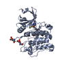
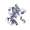
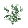
| Title | Crystal structure of MAP4K1 with a SMOL inhibitor |
|---|
 Components Components | Mitogen-activated protein kinase kinase kinase kinase 1 |
|---|
 Keywords Keywords | TRANSFERASE / kinase / inhibitor / drug discovery |
|---|
| Function / homology |  Function and homology information Function and homology information
MAP kinase kinase kinase kinase activity / cellular response to phorbol 13-acetate 12-myristate / JNK cascade / peptidyl-serine phosphorylation / protein autophosphorylation / protein phosphorylation / non-specific serine/threonine protein kinase / cell population proliferation / positive regulation of MAPK cascade / intracellular signal transduction ...MAP kinase kinase kinase kinase activity / cellular response to phorbol 13-acetate 12-myristate / JNK cascade / peptidyl-serine phosphorylation / protein autophosphorylation / protein phosphorylation / non-specific serine/threonine protein kinase / cell population proliferation / positive regulation of MAPK cascade / intracellular signal transduction / protein serine kinase activity / protein serine/threonine kinase activity / ATP binding / membrane / cytoplasmSimilarity search - Function Mitogen-activated protein (MAP) kinase kinase kinase kinase / Domain found in NIK1-like kinases, mouse citron and yeast ROM1, ROM2 / CNH domain / Citron homology (CNH) domain / Citron homology (CNH) domain profile. / : / Protein kinase domain / Serine/Threonine protein kinases, catalytic domain / Protein kinase, ATP binding site / Protein kinases ATP-binding region signature. ...Mitogen-activated protein (MAP) kinase kinase kinase kinase / Domain found in NIK1-like kinases, mouse citron and yeast ROM1, ROM2 / CNH domain / Citron homology (CNH) domain / Citron homology (CNH) domain profile. / : / Protein kinase domain / Serine/Threonine protein kinases, catalytic domain / Protein kinase, ATP binding site / Protein kinases ATP-binding region signature. / Protein kinase domain profile. / Protein kinase domain / Protein kinase-like domain superfamilySimilarity search - Domain/homology |
|---|
| Biological species |  Homo sapiens (human) Homo sapiens (human) |
|---|
| Method |  X-RAY DIFFRACTION / SYNCHROTRON / MOLECULAR REPLACEMENT / Resolution: 2.7 Å X-RAY DIFFRACTION / SYNCHROTRON / MOLECULAR REPLACEMENT / Resolution: 2.7 Å |
|---|
 Authors Authors | Friberg, A. |
|---|
| Funding support | 1items | Organization | Grant number | Country |
|---|
| Not funded | | |
|
|---|
 Citation Citation | Journal: J.Med.Chem. / Year: 2024
Title: Discovery of BAY-405: An Azaindole-Based MAP4K1 Inhibitor for the Enhancement of T-Cell Immunity against Cancer.
Authors: Mowat, J. / Carretero, R. / Leder, G. / Aiguabella Font, N. / Neuhaus, R. / Berndt, S. / Gunther, J. / Friberg, A. / Schafer, M. / Briem, H. / Raschke, M. / Miyatake Ondozabal, H. / ...Authors: Mowat, J. / Carretero, R. / Leder, G. / Aiguabella Font, N. / Neuhaus, R. / Berndt, S. / Gunther, J. / Friberg, A. / Schafer, M. / Briem, H. / Raschke, M. / Miyatake Ondozabal, H. / Buchmann, B. / Boemer, U. / Kreft, B. / Hartung, I.V. / Offringa, R. |
|---|
| History | | Deposition | Jun 8, 2023 | Deposition site: PDBE / Processing site: PDBE |
|---|
| Revision 1.0 | Jun 26, 2024 | Provider: repository / Type: Initial release |
|---|
| Revision 1.1 | Jan 29, 2025 | Group: Database references / Structure summary / Category: citation / citation_author / pdbx_entry_details
Item: _citation.country / _citation.journal_abbrev ..._citation.country / _citation.journal_abbrev / _citation.journal_id_ASTM / _citation.journal_id_CSD / _citation.journal_id_ISSN / _citation.journal_volume / _citation.page_first / _citation.page_last / _citation.pdbx_database_id_DOI / _citation.pdbx_database_id_PubMed / _citation.title / _citation.year / _pdbx_entry_details.has_protein_modification |
|---|
|
|---|
 Movie
Movie Controller


 Open data
 Basic information
 Structure visualization
 Downloads & links
 Other downloads
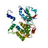
 PDBj
PDBj Assembly
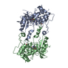

 Spodoptera frugiperda (fall armyworm)
 Sample preparation
 / Beamline: X10SA / Wavelength: 0.99998 Å
 Processing