 Movie
Movie Controller

[English] 日本語
 Yorodumi
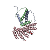
- PDB-8p9e: Crystal structure of wild type p63-p73 heterotetramer (tetrameris... -
+ Open data

- Basic information
| Entry | Database: PDB / ID: 8p9e | ||||||
|---|---|---|---|---|---|---|---|
| Title | Crystal structure of wild type p63-p73 heterotetramer (tetramerisation domain) in complex with darpin 1810 F11 | ||||||
 Components Components |
| ||||||
 Keywords Keywords | DNA BINDING PROTEIN / p63 / p73 / tetramerization domain / darpin / heterotetramer / Structural Genomics / Structural Genomics Consortium / SGC | ||||||
| Function / homology |  Function and homology information Function and homology informationectoderm and mesoderm interaction / epidermal cell division / cloacal septation / positive regulation of somatic stem cell population maintenance / positive regulation of lung ciliated cell differentiation / negative regulation of mesoderm development / prostatic bud formation / cerebrospinal fluid secretion / female genitalia morphogenesis / positive regulation of keratinocyte proliferation ...ectoderm and mesoderm interaction / epidermal cell division / cloacal septation / positive regulation of somatic stem cell population maintenance / positive regulation of lung ciliated cell differentiation / negative regulation of mesoderm development / prostatic bud formation / cerebrospinal fluid secretion / female genitalia morphogenesis / positive regulation of keratinocyte proliferation / establishment of planar polarity / squamous basal epithelial stem cell differentiation involved in prostate gland acinus development / negative regulation of keratinocyte differentiation / polarized epithelial cell differentiation / proximal/distal pattern formation / positive regulation of fibroblast apoptotic process / negative regulation of cardiac muscle cell proliferation / skin morphogenesis / positive regulation of cell cycle G1/S phase transition / negative regulation of intracellular estrogen receptor signaling pathway / sympathetic nervous system development / cranial skeletal system development / post-anal tail morphogenesis / positive regulation of intrinsic apoptotic signaling pathway in response to DNA damage by p53 class mediator / embryonic forelimb morphogenesis / Differentiation of Keratinocytes in Interfollicular Epidermis in Mammalian Skin / embryonic hindlimb morphogenesis / TP53 Regulates Transcription of Death Receptors and Ligands / Activation of PUMA and translocation to mitochondria / Regulation of TP53 Activity through Association with Co-factors / hair follicle morphogenesis / WW domain binding / epithelial cell development / negative regulation of neuron differentiation / TP53 Regulates Transcription of Caspase Activators and Caspases / digestive tract morphogenesis / positive regulation of Notch signaling pathway / regulation of epidermal cell division / positive regulation of stem cell proliferation / odontogenesis of dentin-containing tooth / TP53 Regulates Transcription of Genes Involved in Cytochrome C Release / TP53 regulates transcription of several additional cell death genes whose specific roles in p53-dependent apoptosis remain uncertain / positive regulation of oligodendrocyte differentiation / negative regulation of cellular senescence / keratinocyte proliferation / intrinsic apoptotic signaling pathway in response to DNA damage by p53 class mediator / positive regulation of cell size / Pyroptosis / establishment of skin barrier / positive regulation of osteoblast differentiation / neuron development / mismatch repair / keratinocyte differentiation / Notch signaling pathway / MDM2/MDM4 family protein binding / regulation of mitotic cell cycle / release of cytochrome c from mitochondria / positive regulation of apoptotic signaling pathway / transcription corepressor binding / stem cell proliferation / skeletal system development / determination of adult lifespan / post-embryonic development / TP53 Regulates Metabolic Genes / RNA polymerase II transcription regulatory region sequence-specific DNA binding / hippocampus development / protein tetramerization / promoter-specific chromatin binding / kidney development / intrinsic apoptotic signaling pathway in response to DNA damage / p53 binding / cell junction / cellular senescence / RUNX1 regulates transcription of genes involved in differentiation of HSCs / regulation of gene expression / DNA-binding transcription activator activity, RNA polymerase II-specific / neuron apoptotic process / spermatogenesis / DNA-binding transcription factor binding / negative regulation of neuron apoptotic process / damaged DNA binding / RNA polymerase II-specific DNA-binding transcription factor binding / transcription by RNA polymerase II / DNA-binding transcription factor activity, RNA polymerase II-specific / regulation of cell cycle / transcription cis-regulatory region binding / positive regulation of MAPK cascade / ciliary basal body / RNA polymerase II cis-regulatory region sequence-specific DNA binding / chromatin remodeling / response to xenobiotic stimulus / DNA-binding transcription factor activity / inflammatory response / negative regulation of cell population proliferation / negative regulation of DNA-templated transcription / intracellular membrane-bounded organelle / apoptotic process / DNA damage response / dendrite / centrosome Similarity search - Function | ||||||
| Biological species |  Homo sapiens (human) Homo sapiens (human) | ||||||
| Method |  X-RAY DIFFRACTION / SYNCHROTRON / MOLECULAR REPLACEMENT / Resolution: 2.25 Å X-RAY DIFFRACTION / SYNCHROTRON / MOLECULAR REPLACEMENT / Resolution: 2.25 Å | ||||||
 Authors Authors | Chaikuad, A. / Strubel, A. / Doetsch, V. / Knapp, S. / Structural Genomics Consortium (SGC) | ||||||
| Funding support | 1items
| ||||||
 Citation Citation | Journal: Cell Death Dis / Year: 2023 Title: DARPins detect the formation of hetero-tetramers of p63 and p73 in epithelial tissues and in squamous cell carcinoma. Authors: Strubel, A. / Munick, P. / Hartmann, O. / Chaikuad, A. / Dreier, B. / Schaefer, J.V. / Gebel, J. / Osterburg, C. / Tuppi, M. / Schafer, B. / Buck, V. / Rosenfeldt, M. / Knapp, S. / ...Authors: Strubel, A. / Munick, P. / Hartmann, O. / Chaikuad, A. / Dreier, B. / Schaefer, J.V. / Gebel, J. / Osterburg, C. / Tuppi, M. / Schafer, B. / Buck, V. / Rosenfeldt, M. / Knapp, S. / Pluckthun, A. / Diefenbacher, M.E. / Dotsch, V. #1: Journal: Chemmedchem / Year: 2023Title: Structural Basis of Saccharin Derivative Inhibition of Carbonic Anhydrase IX. Authors: Leitans, J. / Kazaks, A. / Bogans, J. / Supuran, C.T. / Akopjana, I. / Ivanova, J. / Zalubovskis, R. / Tars, K. | ||||||
| History |
|
- Structure visualization
| Structure viewer | Molecule: MolmilJmol/JSmol |
|---|
- Downloads & links
-Download
| PDBx/mmCIF format | 8p9e.cif.gz | 118.8 KB | Display | PDBx/mmCIF format |
|---|---|---|---|---|
| PDB format | pdb8p9e.ent.gz | 91 KB | Display | PDB format |
| PDBx/mmJSON format | 8p9e.json.gz | Tree view | PDBx/mmJSON format | |
| Others |  Other downloads Other downloads |
-Validation report
| Summary document | 8p9e_validation.pdf.gz | 438.8 KB | Display | wwPDB validaton report |
|---|---|---|---|---|
| Full document | 8p9e_full_validation.pdf.gz | 439.5 KB | Display | |
| Data in XML | 8p9e_validation.xml.gz | 11.4 KB | Display | |
| Data in CIF | 8p9e_validation.cif.gz | 14.7 KB | Display | |
| Arichive directory | https://data.pdbj.org/pub/pdb/validation_reports/p9/8p9eftp://data.pdbj.org/pub/pdb/validation_reports/p9/8p9e | HTTPS FTP |
-Related structure data
-Links
 PDBj
PDBj


- Assembly
| Deposited unit | 
| ||||||||
|---|---|---|---|---|---|---|---|---|---|
| 1 | 
| ||||||||
| Unit cell |
|
-Components
| #1: Protein | Mass: 7380.309 Da / Num. of mol.: 1 Source method: isolated from a genetically manipulated source Source: (gene. exp.) Homo sapiens (human) / Gene: TP63, KET, P63, P73H, P73L, TP73L / Production host:  |
|---|---|
| #2: Protein/peptide | Mass: 6034.828 Da / Num. of mol.: 1 Source method: isolated from a genetically manipulated source Source: (gene. exp.) Homo sapiens (human) / Gene: TP73, P73 / Production host: |
| #3: Protein | Mass: 16872.984 Da / Num. of mol.: 1 Source method: isolated from a genetically manipulated source Source: (gene. exp.) |
| #4: Chemical | ChemComp-GOL /   Mass: 92.094 Da / Num. of mol.: 1 / Source method: obtained synthetically / Formula: C3H8O3 Mass: 92.094 Da / Num. of mol.: 1 / Source method: obtained synthetically / Formula: C3H8O3 |
| #5: Water | ChemComp-HOH /  Mass: 18.015 Da / Num. of mol.: 29 / Source method: isolated from a natural source / Formula: H2O Mass: 18.015 Da / Num. of mol.: 29 / Source method: isolated from a natural source / Formula: H2O |
| Has ligand of interest | N |
-Experimental details
-Experiment
| Experiment | Method: X-RAY DIFFRACTION / Number of used crystals: 1 |
|---|
- Sample preparation
| Crystal | Density Matthews: 2.25 Å3/Da / Density % sol: 45.25 % |
|---|---|
| Crystal grow | Temperature: 293.15 K / Method: vapor diffusion, sitting drop / pH: 6.5 Details: 0.2M sodium chloride, 25% PEG3350, 0.1M bis-tris pH 6.5 |
-Data collection
| Diffraction | Mean temperature: 100 K / Serial crystal experiment: N |
|---|---|
| Diffraction source | Source: SYNCHROTRON / Site: SLS  / Beamline: X06SA / Wavelength: 1 Å / Beamline: X06SA / Wavelength: 1 Å |
| Detector | Type: DECTRIS EIGER X 16M / Detector: PIXEL / Date: May 16, 2019 |
| Radiation | Protocol: SINGLE WAVELENGTH / Monochromatic (M) / Laue (L): M / Scattering type: x-ray |
| Radiation wavelength | Wavelength: 1 Å / Relative weight: 1 |
| Reflection | Resolution: 2.25→49.05 Å / Num. obs: 12746 / % possible obs: 99.4 % / Redundancy: 7 % / CC1/2: 0.998 / Rmerge(I) obs: 0.099 / Rpim(I) all: 0.042 / Rrim(I) all: 0.113 / Χ2: 0.87 / Net I/σ(I): 8.6 |
| Reflection shell | Resolution: 2.25→2.33 Å / Redundancy: 6.8 % / Rmerge(I) obs: 0.777 / Mean I/σ(I) obs: 2.1 / Num. unique obs: 1208 / CC1/2: 0.883 / Rpim(I) all: 0.344 / Rrim(I) all: 0.905 / Χ2: 0.86 |
- Processing
| Software |
| ||||||||||||||||||||||||||||||||||||||||||||||||||||||||||||||||||||||||||||||||||||||||||||||||||||||||||||||||||||||||||||||||||||||||||||||||||||||||||||||||||||||||||||||||||||||
|---|---|---|---|---|---|---|---|---|---|---|---|---|---|---|---|---|---|---|---|---|---|---|---|---|---|---|---|---|---|---|---|---|---|---|---|---|---|---|---|---|---|---|---|---|---|---|---|---|---|---|---|---|---|---|---|---|---|---|---|---|---|---|---|---|---|---|---|---|---|---|---|---|---|---|---|---|---|---|---|---|---|---|---|---|---|---|---|---|---|---|---|---|---|---|---|---|---|---|---|---|---|---|---|---|---|---|---|---|---|---|---|---|---|---|---|---|---|---|---|---|---|---|---|---|---|---|---|---|---|---|---|---|---|---|---|---|---|---|---|---|---|---|---|---|---|---|---|---|---|---|---|---|---|---|---|---|---|---|---|---|---|---|---|---|---|---|---|---|---|---|---|---|---|---|---|---|---|---|---|---|---|---|---|
| Refinement | Method to determine structure: MOLECULAR REPLACEMENT / Resolution: 2.25→49.05 Å / Cor.coef. Fo:Fc: 0.966 / Cor.coef. Fo:Fc free: 0.926 / SU B: 20.913 / SU ML: 0.225 / Cross valid method: THROUGHOUT / ESU R: 0.328 / ESU R Free: 0.239 / Stereochemistry target values: MAXIMUM LIKELIHOOD / Details: HYDROGENS HAVE BEEN ADDED IN THE RIDING POSITIONS
| ||||||||||||||||||||||||||||||||||||||||||||||||||||||||||||||||||||||||||||||||||||||||||||||||||||||||||||||||||||||||||||||||||||||||||||||||||||||||||||||||||||||||||||||||||||||
| Solvent computation | Ion probe radii: 0.8 Å / Shrinkage radii: 0.8 Å / VDW probe radii: 1.2 Å / Solvent model: MASK | ||||||||||||||||||||||||||||||||||||||||||||||||||||||||||||||||||||||||||||||||||||||||||||||||||||||||||||||||||||||||||||||||||||||||||||||||||||||||||||||||||||||||||||||||||||||
| Displacement parameters | Biso mean: 61.647 Å2
| ||||||||||||||||||||||||||||||||||||||||||||||||||||||||||||||||||||||||||||||||||||||||||||||||||||||||||||||||||||||||||||||||||||||||||||||||||||||||||||||||||||||||||||||||||||||
| Refinement step | Cycle: 1 / Resolution: 2.25→49.05 Å
| ||||||||||||||||||||||||||||||||||||||||||||||||||||||||||||||||||||||||||||||||||||||||||||||||||||||||||||||||||||||||||||||||||||||||||||||||||||||||||||||||||||||||||||||||||||||
| Refine LS restraints |
|