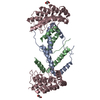
Entry Database : PDB / ID : 8p9dTitle Crystal structure of p63-p73 heterotetramer (tetramerisation domain) in complex with darpin 1810 A2 Darpin 1810 A2 Tumor protein 63 Tumor protein p73 Keywords / / / / / / / / Function / homology Function Domain/homology Component
/ / / / / / / / / / / / / / / / / / / / / / / / / / / / / / / / / / / / / / / / / / / / / / / / / / / / / / / / / / / / / / / / / / / / / / / / / / / / / / / / / / / / / / / / / / / / / / / / / / / / / / / / / / / / / / / / / / / / / / / / / / / / / / / / / / / Biological species Homo sapiens (human)Lama glama (llama)Method / / / Resolution : 2.7 Å Authors Chaikuad, A. / Strubel, A. / Doetsch, V. / Knapp, S. / Structural Genomics Consortium (SGC) Funding support 1items Organization Grant number Country Not funded
Journal : Cell Death Dis / Year : 2023Title : DARPins detect the formation of hetero-tetramers of p63 and p73 in epithelial tissues and in squamous cell carcinoma.Authors: Strubel, A. / Munick, P. / Hartmann, O. / Chaikuad, A. / Dreier, B. / Schaefer, J.V. / Gebel, J. / Osterburg, C. / Tuppi, M. / Schafer, B. / Buck, V. / Rosenfeldt, M. / Knapp, S. / ... Authors : Strubel, A. / Munick, P. / Hartmann, O. / Chaikuad, A. / Dreier, B. / Schaefer, J.V. / Gebel, J. / Osterburg, C. / Tuppi, M. / Schafer, B. / Buck, V. / Rosenfeldt, M. / Knapp, S. / Pluckthun, A. / Diefenbacher, M.E. / Dotsch, V. History Deposition Jun 5, 2023 Deposition site / Processing site Revision 1.0 Nov 8, 2023 Provider / Type Revision 1.1 Nov 15, 2023 Group Database references / Derived calculations ... Database references / Derived calculations / Source and taxonomy / Structure summary Category citation / entity ... citation / entity / entity_src_gen / pdbx_entity_src_syn / pdbx_struct_assembly Item _citation.journal_id_ISSN / _entity.pdbx_description ... _citation.journal_id_ISSN / _entity.pdbx_description / _entity.src_method / _pdbx_struct_assembly.details
Show all Show less
 Movie
Movie Controller

 Yorodumi
 Open data
 Basic information
 Components
 Keywords
 Function and homology information
 Homo sapiens (human)
 X-RAY DIFFRACTION /
  Authors
 Citation
 Structure visualization
 Downloads & links
 Other downloads
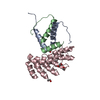
 PDBj
PDBj


 Assembly

 Mass: 18.015 Da / Num. of mol.: 6 / Source method: isolated from a natural source / Formula: H2O
 Sample preparation
 / Beamline: X06SA / Wavelength: 1 Å
 Processing