| Entry | Database: PDB / ID: 8p6j
|
|---|
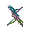
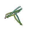
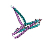
| Title | Structure of the hypervariable region of Streptococcus pyogenes M3 protein in complex with a collagen peptide |
|---|
 Components Components | - Antiphagocytic M protein, type 3
- collagen II-27 Toolkit peptide (JDM238)
|
|---|
 Keywords Keywords | PROTEIN BINDING / M protein / Streptococcus pyogenes / hypervariable region / collagen-binding domain |
|---|
| Function / homology |  Function and homology information Function and homology information
: / Streptococcal M proteins D repeats region profile. / : / Streptococcal M proteins C repeat profile. / M protein-type anchor domain / YSIRK type signal peptide / YSIRK Gram-positive signal peptide / LPXTG cell wall anchor motif / Gram-positive cocci surface proteins LPxTG motif profile. / LPXTG cell wall anchor domainSimilarity search - Domain/homology |
|---|
| Biological species |  Streptococcus pyogenes serotype M3 (bacteria) Streptococcus pyogenes serotype M3 (bacteria)
 Homo sapiens (human) Homo sapiens (human) |
|---|
| Method |  X-RAY DIFFRACTION / SYNCHROTRON / MOLECULAR REPLACEMENT / Resolution: 2.324 Å X-RAY DIFFRACTION / SYNCHROTRON / MOLECULAR REPLACEMENT / Resolution: 2.324 Å |
|---|
 Authors Authors | Wojnowska, M. / Schwarz-Linek, U. |
|---|
| Funding support |  United Kingdom, 1items United Kingdom, 1items | Organization | Grant number | Country |
|---|
| Medical Research Council (MRC, United Kingdom) | MR/N009681/1 | United Kingdom |
|
|---|
 Citation Citation | Journal: Biorxiv / Year: 2024
Title: Structural basis for collagen recognition by the Streptococcus pyogenes M3 protein and its involvement in biofilm
Authors: Wojnowska, M. / Wajima, T. / Yelland, T. / Ludewig, H. / Hagan, R.M. / Watt, G. / Hamaia, S.W. / Bihan, D. / Malcor, J.D. / Bonna, A. / Bergsten, H. / Svensson, M. / Oppegaard, O. / Skrede, ...Authors: Wojnowska, M. / Wajima, T. / Yelland, T. / Ludewig, H. / Hagan, R.M. / Watt, G. / Hamaia, S.W. / Bihan, D. / Malcor, J.D. / Bonna, A. / Bergsten, H. / Svensson, M. / Oppegaard, O. / Skrede, S. / Arnell, P. / Hyldegaard, O. / Farndale, R.W. / Norrby-Teglund, A. / Schwarz-Linek, U. |
|---|
| History | | Deposition | May 26, 2023 | Deposition site: PDBE / Processing site: PDBE |
|---|
| Revision 1.0 | Jun 21, 2023 | Provider: repository / Type: Initial release |
|---|
| Revision 1.1 | Sep 13, 2023 | Group: Data collection / Database references ...Data collection / Database references / Derived calculations / Refinement description / Source and taxonomy / Structure summary
Category: chem_comp_atom / chem_comp_bond ...chem_comp_atom / chem_comp_bond / entity / entity_src_gen / pdbx_struct_assembly / pdbx_struct_assembly_gen / pdbx_struct_assembly_prop / pdbx_struct_oper_list / struct / struct_ncs_dom_lim / struct_ref / struct_ref_seq / struct_ref_seq_dif
Item: _entity.pdbx_description / _entity_src_gen.pdbx_gene_src_gene ..._entity.pdbx_description / _entity_src_gen.pdbx_gene_src_gene / _struct.title / _struct_ncs_dom_lim.beg_auth_comp_id / _struct_ncs_dom_lim.beg_label_asym_id / _struct_ncs_dom_lim.beg_label_comp_id / _struct_ncs_dom_lim.beg_label_seq_id / _struct_ncs_dom_lim.end_auth_comp_id / _struct_ncs_dom_lim.end_label_asym_id / _struct_ncs_dom_lim.end_label_comp_id / _struct_ncs_dom_lim.end_label_seq_id / _struct_ref.db_code / _struct_ref.pdbx_db_accession / _struct_ref_seq.pdbx_db_accession / _struct_ref_seq_dif.pdbx_seq_db_accession_code |
|---|
| Revision 1.2 | Jan 15, 2025 | Group: Database references / Structure summary
Category: citation / citation_author ...citation / citation_author / pdbx_entry_details / pdbx_modification_feature
Item: _citation.country / _citation.journal_abbrev ..._citation.country / _citation.journal_abbrev / _citation.journal_id_CSD / _citation.journal_id_ISSN / _citation.pdbx_database_id_DOI / _citation.title / _citation.year / _pdbx_entry_details.has_protein_modification |
|---|
|
|---|
 Movie
Movie Controller

 Yorodumi
 Open data
 Basic information
 Structure visualization
 Downloads & links
 Other downloads
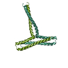
 PDBj
PDBj Assembly