 Movie
Movie Controller

[English] 日本語
 Yorodumi
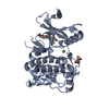
- PDB-8p5k: Kinase domain of mutant human ULK1 in complex with compound MRT68921 -
+ Open data

- Basic information
| Entry | Database: PDB / ID: 8p5k | ||||||
|---|---|---|---|---|---|---|---|
| Title | Kinase domain of mutant human ULK1 in complex with compound MRT68921 | ||||||
 Components Components | Serine/threonine-protein kinase ULK1 | ||||||
 Keywords Keywords | TRANSFERASE / Autophagy / Unc-like 1 kinase | ||||||
| Function / homology |  Function and homology information Function and homology informationneuron projection regeneration / omegasome membrane / negative regulation of collateral sprouting / Atg1/ULK1 kinase complex / positive regulation of autophagosome assembly / phagophore assembly site membrane / piecemeal microautophagy of the nucleus / RAB GEFs exchange GTP for GDP on RABs / regulation of tumor necrosis factor-mediated signaling pathway / axon extension ...neuron projection regeneration / omegasome membrane / negative regulation of collateral sprouting / Atg1/ULK1 kinase complex / positive regulation of autophagosome assembly / phagophore assembly site membrane / piecemeal microautophagy of the nucleus / RAB GEFs exchange GTP for GDP on RABs / regulation of tumor necrosis factor-mediated signaling pathway / axon extension / phagophore assembly site / TBC/RABGAPs / reticulophagy / response to starvation / Receptor Mediated Mitophagy / cellular response to stress / Macroautophagy / autophagosome membrane / regulation of macroautophagy / negative regulation of protein-containing complex assembly / autophagosome assembly / mitophagy / positive regulation of autophagy / cellular response to nutrient levels / autophagosome / peptidyl-serine phosphorylation / regulation of autophagy / macroautophagy / Regulation of TNFR1 signaling / recycling endosome / autophagy / small GTPase binding / neuron projection development / protein autophosphorylation / intracellular protein localization / GTPase binding / protein phosphorylation / mitochondrial outer membrane / non-specific serine/threonine protein kinase / negative regulation of cell population proliferation / protein serine kinase activity / axon / protein serine/threonine kinase activity / endoplasmic reticulum membrane / protein-containing complex binding / signal transduction / ATP binding / identical protein binding / cytoplasm / cytosol Similarity search - Function | ||||||
| Biological species |  Homo sapiens (human) Homo sapiens (human) | ||||||
| Method |  X-RAY DIFFRACTION / SYNCHROTRON / MOLECULAR REPLACEMENT / Resolution: 2.209 Å X-RAY DIFFRACTION / SYNCHROTRON / MOLECULAR REPLACEMENT / Resolution: 2.209 Å | ||||||
 Authors Authors | Battista, T. / Semrau, M.S. / Heroux, A. / Lolli, G. / Storici, P. | ||||||
| Funding support | 1items
| ||||||
 Citation Citation | Journal: To Be Published Title: Crystal structures of ULK1 in complex with KCGS compounds Authors: Battista, T. / Semrau, M.S. / Heroux, A. / Lolli, G. / Storici, P. | ||||||
| History |
|
- Structure visualization
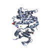
| Structure viewer | Molecule: MolmilJmol/JSmol |
|---|
- Downloads & links
-Download
| PDBx/mmCIF format | 8p5k.cif.gz | 458.8 KB | Display | PDBx/mmCIF format |
|---|---|---|---|---|
| PDB format | pdb8p5k.ent.gz | 371.2 KB | Display | PDB format |
| PDBx/mmJSON format | 8p5k.json.gz | Tree view | PDBx/mmJSON format | |
| Others |  Other downloads Other downloads |
-Validation report
| Arichive directory | https://data.pdbj.org/pub/pdb/validation_reports/p5/8p5kftp://data.pdbj.org/pub/pdb/validation_reports/p5/8p5k | HTTPS FTP |
|---|
-Related structure data


-Links
 PDBj
PDBj


- Assembly
| Deposited unit | 
| ||||||||||||||||||||||||||||||||||||||||||||||||||||||||||||||||||||||||||||||||||||||||||||||||||||||||||||||||||||||||||||||||||||||||||||||||
|---|---|---|---|---|---|---|---|---|---|---|---|---|---|---|---|---|---|---|---|---|---|---|---|---|---|---|---|---|---|---|---|---|---|---|---|---|---|---|---|---|---|---|---|---|---|---|---|---|---|---|---|---|---|---|---|---|---|---|---|---|---|---|---|---|---|---|---|---|---|---|---|---|---|---|---|---|---|---|---|---|---|---|---|---|---|---|---|---|---|---|---|---|---|---|---|---|---|---|---|---|---|---|---|---|---|---|---|---|---|---|---|---|---|---|---|---|---|---|---|---|---|---|---|---|---|---|---|---|---|---|---|---|---|---|---|---|---|---|---|---|---|---|---|---|---|
| 1 | 
| ||||||||||||||||||||||||||||||||||||||||||||||||||||||||||||||||||||||||||||||||||||||||||||||||||||||||||||||||||||||||||||||||||||||||||||||||
| 2 | 
| ||||||||||||||||||||||||||||||||||||||||||||||||||||||||||||||||||||||||||||||||||||||||||||||||||||||||||||||||||||||||||||||||||||||||||||||||
| 3 | 
| ||||||||||||||||||||||||||||||||||||||||||||||||||||||||||||||||||||||||||||||||||||||||||||||||||||||||||||||||||||||||||||||||||||||||||||||||
| 4 | 
| ||||||||||||||||||||||||||||||||||||||||||||||||||||||||||||||||||||||||||||||||||||||||||||||||||||||||||||||||||||||||||||||||||||||||||||||||
| Unit cell |
| ||||||||||||||||||||||||||||||||||||||||||||||||||||||||||||||||||||||||||||||||||||||||||||||||||||||||||||||||||||||||||||||||||||||||||||||||
| Noncrystallographic symmetry (NCS) | NCS domain:
NCS domain segments: Beg auth comp-ID: THR / Beg label comp-ID: THR / Auth asym-ID: A / Label asym-ID: A
NCS ensembles :
|
-Components
-Protein , 1 types, 4 molecules ABCD
| #1: Protein | Mass: 32121.098 Da / Num. of mol.: 4 / Mutation: R245A, E246A Source method: isolated from a genetically manipulated source Source: (gene. exp.) Homo sapiens (human) / Gene: ULK1, KIAA0722 / Production host:  References: UniProt: O75385, non-specific serine/threonine protein kinase |
|---|
-Non-polymers , 5 types, 634 molecules 


| #2: Chemical | ChemComp-HVH / ~{  Mass: 434.577 Da / Num. of mol.: 4 / Source method: obtained synthetically / Formula: C25H34N6O / Feature type: SUBJECT OF INVESTIGATION Mass: 434.577 Da / Num. of mol.: 4 / Source method: obtained synthetically / Formula: C25H34N6O / Feature type: SUBJECT OF INVESTIGATION#3: Chemical |  Mass: 92.094 Da / Num. of mol.: 3 / Source method: obtained synthetically / Formula: C3H8O3 Mass: 92.094 Da / Num. of mol.: 3 / Source method: obtained synthetically / Formula: C3H8O3#4: Chemical | ChemComp-MG /  Mass: 24.305 Da / Num. of mol.: 8 / Source method: obtained synthetically / Formula: Mg Mass: 24.305 Da / Num. of mol.: 8 / Source method: obtained synthetically / Formula: Mg#5: Chemical |  Mass: 94.971 Da / Num. of mol.: 2 / Source method: obtained synthetically / Formula: PO4 Mass: 94.971 Da / Num. of mol.: 2 / Source method: obtained synthetically / Formula: PO4#6: Water | ChemComp-HOH / | Mass: 18.015 Da / Num. of mol.: 617 / Source method: isolated from a natural source / Formula: H2O |
|---|
-Details
| Has ligand of interest | Y |
|---|---|
| Has protein modification | Y |
-Experimental details
-Experiment
| Experiment | Method: X-RAY DIFFRACTION / Number of used crystals: 1 |
|---|
- Sample preparation
| Crystal | Density Matthews: 3.5 Å3/Da / Density % sol: 64.88 % |
|---|---|
| Crystal grow | Temperature: 277.15 K / Method: vapor diffusion, hanging drop / pH: 6 / Details: 0.3-0.8 M NaAcetate pH 6, 20-26% w/v PEG3350 |
-Data collection
| Diffraction | Mean temperature: 100 K / Serial crystal experiment: N |
|---|---|
| Diffraction source | Source: SYNCHROTRON / Site: ELETTRA  / Beamline: 11.2C / Wavelength: 1 Å / Beamline: 11.2C / Wavelength: 1 Å |
| Detector | Type: DECTRIS PILATUS 6M / Detector: PIXEL / Date: May 5, 2023 |
| Radiation | Protocol: SINGLE WAVELENGTH / Monochromatic (M) / Laue (L): M / Scattering type: x-ray |
| Radiation wavelength | Wavelength: 1 Å / Relative weight: 1 |
| Reflection | Resolution: 2.209→85.075 Å / Num. obs: 88463 / % possible obs: 99.7 % / Redundancy: 6.6 % / CC1/2: 0.999 / Net I/σ(I): 16.6 |
| Reflection shell | Resolution: 2.209→2.247 Å / Mean I/σ(I) obs: 2.1 / Num. unique obs: 4401 / CC1/2: 0.714 / % possible all: 99.2 |
- Processing
| Software |
| ||||||||||||||||||||||||||||||||||||||||||||||||||||||||||||||||||||||||||||||||||||||||||||||||||||||||||||||||||||||||||||||||||||||||||||||||||||||||||||||||||||||||||||||||||||||
|---|---|---|---|---|---|---|---|---|---|---|---|---|---|---|---|---|---|---|---|---|---|---|---|---|---|---|---|---|---|---|---|---|---|---|---|---|---|---|---|---|---|---|---|---|---|---|---|---|---|---|---|---|---|---|---|---|---|---|---|---|---|---|---|---|---|---|---|---|---|---|---|---|---|---|---|---|---|---|---|---|---|---|---|---|---|---|---|---|---|---|---|---|---|---|---|---|---|---|---|---|---|---|---|---|---|---|---|---|---|---|---|---|---|---|---|---|---|---|---|---|---|---|---|---|---|---|---|---|---|---|---|---|---|---|---|---|---|---|---|---|---|---|---|---|---|---|---|---|---|---|---|---|---|---|---|---|---|---|---|---|---|---|---|---|---|---|---|---|---|---|---|---|---|---|---|---|---|---|---|---|---|---|---|
| Refinement | Method to determine structure: MOLECULAR REPLACEMENT / Resolution: 2.209→85.075 Å / Cor.coef. Fo:Fc: 0.961 / Cor.coef. Fo:Fc free: 0.95 / SU B: 4.874 / SU ML: 0.117 / Cross valid method: FREE R-VALUE / ESU R: 0.182 / ESU R Free: 0.155 Details: Hydrogens have been added in their riding positions
| ||||||||||||||||||||||||||||||||||||||||||||||||||||||||||||||||||||||||||||||||||||||||||||||||||||||||||||||||||||||||||||||||||||||||||||||||||||||||||||||||||||||||||||||||||||||
| Solvent computation | Ion probe radii: 0.8 Å / Shrinkage radii: 0.8 Å / VDW probe radii: 1.2 Å / Solvent model: MASK BULK SOLVENT | ||||||||||||||||||||||||||||||||||||||||||||||||||||||||||||||||||||||||||||||||||||||||||||||||||||||||||||||||||||||||||||||||||||||||||||||||||||||||||||||||||||||||||||||||||||||
| Displacement parameters | Biso mean: 40.344 Å2
| ||||||||||||||||||||||||||||||||||||||||||||||||||||||||||||||||||||||||||||||||||||||||||||||||||||||||||||||||||||||||||||||||||||||||||||||||||||||||||||||||||||||||||||||||||||||
| Refinement step | Cycle: LAST / Resolution: 2.209→85.075 Å
| ||||||||||||||||||||||||||||||||||||||||||||||||||||||||||||||||||||||||||||||||||||||||||||||||||||||||||||||||||||||||||||||||||||||||||||||||||||||||||||||||||||||||||||||||||||||
| Refine LS restraints |
|