 Movie
Movie Controller

[English] 日本語
 Yorodumi
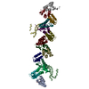
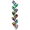
- PDB-8p10: The crystal structure of the C-terminal domain of Mengla nucleoprotein -
+ Open data

- Basic information
| Entry | Database: PDB / ID: 8p10 | ||||||
|---|---|---|---|---|---|---|---|
| Title | The crystal structure of the C-terminal domain of Mengla nucleoprotein | ||||||
 Components Components |
| ||||||
 Keywords Keywords | VIRAL PROTEIN / Nucleoprotein / Filovirus / Mengla | ||||||
| Function / homology | Ebola nucleoprotein / Ebola nucleoprotein / viral RNA genome packaging / helical viral capsid / viral nucleocapsid / host cell cytoplasm / ribonucleoprotein complex / RNA binding / Nucleoprotein Function and homology information Function and homology information | ||||||
| Biological species |  Mengla dianlovirus Mengla dianlovirus | ||||||
| Method |  X-RAY DIFFRACTION / SYNCHROTRON / MOLECULAR REPLACEMENT / Resolution: 3.26 Å X-RAY DIFFRACTION / SYNCHROTRON / MOLECULAR REPLACEMENT / Resolution: 3.26 Å | ||||||
 Authors Authors | Ferrero, D.S. / Tomas Gilabert, O. / Verdaguer, N. | ||||||
| Funding support |  Spain, 1items Spain, 1items
| ||||||
 Citation Citation | Journal: Microbiol Spectr / Year: 2023 Title: Structural insights on the nucleoprotein C-terminal domain of Mengla virus. Authors: Ferrero, D.S. / Tomas Gilabert, O. / Verdaguer, N. | ||||||
| History |
|
- Structure visualization
| Structure viewer | Molecule: MolmilJmol/JSmol |
|---|
- Downloads & links
-Download
| PDBx/mmCIF format | 8p10.cif.gz | 458 KB | Display | PDBx/mmCIF format |
|---|---|---|---|---|
| PDB format | pdb8p10.ent.gz | 316.8 KB | Display | PDB format |
| PDBx/mmJSON format | 8p10.json.gz | Tree view | PDBx/mmJSON format | |
| Others |  Other downloads Other downloads |
-Validation report
| Arichive directory | https://data.pdbj.org/pub/pdb/validation_reports/p1/8p10ftp://data.pdbj.org/pub/pdb/validation_reports/p1/8p10 | HTTPS FTP |
|---|
-Related structure data
-Links
 PDBj
PDBj
- Assembly
| Deposited unit | 
| ||||||||||||
|---|---|---|---|---|---|---|---|---|---|---|---|---|---|
| 1 | 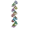
| ||||||||||||
| 2 | 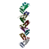
| ||||||||||||
| Unit cell |
|
-Components
| #1: Protein | Mass: 14277.541 Da / Num. of mol.: 28 Source method: isolated from a genetically manipulated source Source: (gene. exp.) Mengla dianlovirus / Gene: NP / Production host:  #2: Protein/peptide | | Mass: 714.767 Da / Num. of mol.: 1 Source method: isolated from a genetically manipulated source Source: (gene. exp.) Mengla dianlovirus / Production host: |
|---|
-Experimental details
-Experiment
| Experiment | Method: X-RAY DIFFRACTION / Number of used crystals: 1 |
|---|
- Sample preparation
| Crystal grow | Temperature: 293 K / Method: vapor diffusion, sitting drop / pH: 6.5 Details: 0.2M Ammonium sulfate, 0.1M MES pH 6.5, 30% PEG5000MME |
|---|
-Data collection
| Diffraction | Mean temperature: 100 K / Serial crystal experiment: N |
|---|---|
| Diffraction source | Source: SYNCHROTRON / Site: Diamond  / Beamline: I03 / Wavelength: 0.9762 Å / Beamline: I03 / Wavelength: 0.9762 Å |
| Detector | Type: DECTRIS EIGER2 XE 16M / Detector: PIXEL / Date: May 20, 2020 |
| Radiation | Protocol: SINGLE WAVELENGTH / Monochromatic (M) / Laue (L): M / Scattering type: x-ray |
| Radiation wavelength | Wavelength: 0.9762 Å / Relative weight: 1 |
| Reflection | Resolution: 3.25→142.156 Å / Num. obs: 30881 / % possible obs: 89.2 % / Redundancy: 3.7 % / Biso Wilson estimate: 104.6 Å2 / CC1/2: 0.998 / Rmerge(I) obs: 0.103 / Net I/σ(I): 8.2 |
| Reflection shell | Resolution: 3.256→4.052 Å / Rmerge(I) obs: 0.713 / Num. unique obs: 6177 / CC1/2: 0.71 |
- Processing
| Software |
| ||||||||||||||||||||||||||||||||||||||||||||||||||||||||||||||||||||||||||||||||||||
|---|---|---|---|---|---|---|---|---|---|---|---|---|---|---|---|---|---|---|---|---|---|---|---|---|---|---|---|---|---|---|---|---|---|---|---|---|---|---|---|---|---|---|---|---|---|---|---|---|---|---|---|---|---|---|---|---|---|---|---|---|---|---|---|---|---|---|---|---|---|---|---|---|---|---|---|---|---|---|---|---|---|---|---|---|---|
| Refinement | Method to determine structure: MOLECULAR REPLACEMENT / Resolution: 3.26→47.99 Å / SU ML: 0.4981 / Cross valid method: FREE R-VALUE / σ(F): 1.96 / Phase error: 37.9231 Stereochemistry target values: GeoStd + Monomer Library + CDL v1.2
| ||||||||||||||||||||||||||||||||||||||||||||||||||||||||||||||||||||||||||||||||||||
| Solvent computation | Shrinkage radii: 0.9 Å / VDW probe radii: 1.1 Å / Solvent model: FLAT BULK SOLVENT MODEL | ||||||||||||||||||||||||||||||||||||||||||||||||||||||||||||||||||||||||||||||||||||
| Displacement parameters | Biso mean: 120.92 Å2 | ||||||||||||||||||||||||||||||||||||||||||||||||||||||||||||||||||||||||||||||||||||
| Refinement step | Cycle: LAST / Resolution: 3.26→47.99 Å
| ||||||||||||||||||||||||||||||||||||||||||||||||||||||||||||||||||||||||||||||||||||
| Refine LS restraints |
| ||||||||||||||||||||||||||||||||||||||||||||||||||||||||||||||||||||||||||||||||||||
| LS refinement shell |
|
