 Movie
Movie Controller

[English] 日本語
 Yorodumi
- PDB-8own: CryoEM structure of glutamate dehydrogenase isoform 2 from Arabid... -
+ Open data

- Basic information
| Entry | Database: PDB / ID: 8own | ||||||
|---|---|---|---|---|---|---|---|
| Title | CryoEM structure of glutamate dehydrogenase isoform 2 from Arabidopsis thaliana in apo-form | ||||||
 Components Components | Glutamate dehydrogenase 2 | ||||||
 Keywords Keywords | OXIDOREDUCTASE / glutamic acid / calcium / NAD cofactor / nitrogen metabolism | ||||||
| Function / homology |  Function and homology information Function and homology informationL-glutamate dehydrogenase [NAD(P)+] activity / glutamate dehydrogenase [NAD(P)+] / L-glutamate dehydrogenase (NADP+) activity / L-glutamate dehydrogenase (NAD+) activity / L-glutamate catabolic process / plant-type vacuole / cobalt ion binding / copper ion binding / mitochondrion / zinc ion binding ...L-glutamate dehydrogenase [NAD(P)+] activity / glutamate dehydrogenase [NAD(P)+] / L-glutamate dehydrogenase (NADP+) activity / L-glutamate dehydrogenase (NAD+) activity / L-glutamate catabolic process / plant-type vacuole / cobalt ion binding / copper ion binding / mitochondrion / zinc ion binding / ATP binding / plasma membrane Similarity search - Function | ||||||
| Biological species |  | ||||||
| Method | ELECTRON MICROSCOPY / single particle reconstruction / cryo EM / Resolution: 3.26 Å | ||||||
 Authors Authors | Grzechowiak, M. / Ruszkowski, M. | ||||||
| Funding support |  Poland, 1items Poland, 1items
| ||||||
 Citation Citation | Journal: Plant Physiol Biochem / Year: 2023 Title: Structural and functional studies of Arabidopsis thaliana glutamate dehydrogenase isoform 2 demonstrate enzyme dynamics and identify its calcium binding site. Authors: Marta Grzechowiak / Joanna Sliwiak / Mariusz Jaskolski / Milosz Ruszkowski / Abstract: Glutamate dehydrogenase (GDH) is an enzyme at the crossroad of plant nitrogen and carbon metabolism. GDH catalyzes the conversion of 2-oxoglutarate into glutamate (2OG → Glu), utilizing ammonia as ...Glutamate dehydrogenase (GDH) is an enzyme at the crossroad of plant nitrogen and carbon metabolism. GDH catalyzes the conversion of 2-oxoglutarate into glutamate (2OG → Glu), utilizing ammonia as cosubstrate and NADH as coenzyme. The GDH reaction is reversible, meaning that the NAD-dependent reaction (Glu → 2OG) releases ammonia. In Arabidopsis thaliana, three GDH isoforms exist, AtGDH1, AtGDH2, and AtGDH3. The subject of this work is AtGDH2. Previous reports have suggested that enzymes homologous to AtGDH2 contain a calcium-binding EF-hand motif located in the coenzyme binding domain. Here, we show that while AtGDH2 indeed does bind calcium, the binding occurs elsewhere and the region predicted to be the EF-hand motif has a completely different structure. As the true calcium binding site is > 20 Å away from the active site, it seems to play a structural, rather than catalytic role. We also performed comparative kinetic characterization of AtGDH1 and AtGDH2 using spectroscopic methods and isothermal titration calorimetry, to note that the isoenzymes generally exhibit similar behavior, with calcium having only a minor effect. However, the spatial and temporal changes in the gene expression profiles of the three AtGDH genes point to AtGDH2 as the most prevalent isoform. | ||||||
| History |
|
- Structure visualization
| Structure viewer | Molecule: MolmilJmol/JSmol |
|---|
- Downloads & links
-Download
| PDBx/mmCIF format | 8own.cif.gz | 414.9 KB | Display | PDBx/mmCIF format |
|---|---|---|---|---|
| PDB format | pdb8own.ent.gz | 336.8 KB | Display | PDB format |
| PDBx/mmJSON format | 8own.json.gz | Tree view | PDBx/mmJSON format | |
| Others |  Other downloads Other downloads |
-Validation report
| Arichive directory | https://data.pdbj.org/pub/pdb/validation_reports/ow/8ownftp://data.pdbj.org/pub/pdb/validation_reports/ow/8own | HTTPS FTP |
|---|
-Related structure data
| Related structure data |  17240MC 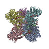 8owmC C: citing same article ( M: map data used to model this data |
|---|---|
| Similar structure data |
-Links
 PDBj
PDBj- Assembly
| Deposited unit | 
| ||||||||||||||||||||||||||||||||||||||||||||||||||||||||||||||||||||||||||||||||||||||||||||||
|---|---|---|---|---|---|---|---|---|---|---|---|---|---|---|---|---|---|---|---|---|---|---|---|---|---|---|---|---|---|---|---|---|---|---|---|---|---|---|---|---|---|---|---|---|---|---|---|---|---|---|---|---|---|---|---|---|---|---|---|---|---|---|---|---|---|---|---|---|---|---|---|---|---|---|---|---|---|---|---|---|---|---|---|---|---|---|---|---|---|---|---|---|---|---|---|
| 1 |
| ||||||||||||||||||||||||||||||||||||||||||||||||||||||||||||||||||||||||||||||||||||||||||||||
| Noncrystallographic symmetry (NCS) | NCS domain:
NCS domain segments:
NCS oper:
|
-Components
| #1: Protein | Mass: 45027.188 Da / Num. of mol.: 6 Source method: isolated from a genetically manipulated source Source: (gene. exp.)  References: UniProt: Q38946, glutamate dehydrogenase [NAD(P)+] #2: Chemical | ChemComp-CA /   Mass: 40.078 Da / Num. of mol.: 6 / Source method: obtained synthetically / Formula: Ca / Feature type: SUBJECT OF INVESTIGATION Mass: 40.078 Da / Num. of mol.: 6 / Source method: obtained synthetically / Formula: Ca / Feature type: SUBJECT OF INVESTIGATIONHas ligand of interest | Y | |
|---|
-Experimental details
-Experiment
| Experiment | Method: ELECTRON MICROSCOPY |
|---|---|
| EM experiment | Aggregation state: PARTICLE / 3D reconstruction method: single particle reconstruction |
- Sample preparation
| Component | Name: homohexameric Arabidopsis thaliana glutamate dehydrogenase isoform 2 Type: ORGANELLE OR CELLULAR COMPONENT / Entity ID: #1 / Source: RECOMBINANT | ||||||||||||||||||||||||||||||
|---|---|---|---|---|---|---|---|---|---|---|---|---|---|---|---|---|---|---|---|---|---|---|---|---|---|---|---|---|---|---|---|
| Molecular weight | Value: 0.27 MDa / Experimental value: NO | ||||||||||||||||||||||||||||||
| Source (natural) | Organism: | ||||||||||||||||||||||||||||||
| Source (recombinant) | Organism: | ||||||||||||||||||||||||||||||
| Buffer solution | pH: 7.5 | ||||||||||||||||||||||||||||||
| Buffer component |
| ||||||||||||||||||||||||||||||
| Specimen | Conc.: 1 mg/ml / Embedding applied: NO / Shadowing applied: NO / Staining applied: NO / Vitrification applied: YES | ||||||||||||||||||||||||||||||
| Specimen support | Grid material: COPPER / Grid type: Quantifoil R1.2/1.3 | ||||||||||||||||||||||||||||||
| Vitrification | Instrument: FEI VITROBOT MARK IV / Cryogen name: ETHANE / Humidity: 100 % / Chamber temperature: 277 K |
- Electron microscopy imaging
| Experimental equipment |  Model: Titan Krios / Image courtesy: FEI Company |
|---|---|
| Microscopy | Model: FEI TITAN KRIOS |
| Electron gun | Electron source:  FIELD EMISSION GUN / Accelerating voltage: 300 kV / Illumination mode: FLOOD BEAM FIELD EMISSION GUN / Accelerating voltage: 300 kV / Illumination mode: FLOOD BEAM |
| Electron lens | Mode: BRIGHT FIELD / Nominal defocus max: 3500 nm / Nominal defocus min: 500 nm / Calibrated defocus min: 231 nm / Calibrated defocus max: 4623 nm / Cs: 2.7 mm |
| Specimen holder | Specimen holder model: FEI TITAN KRIOS AUTOGRID HOLDER |
| Image recording | Electron dose: 40 e/Å2 / Film or detector model: FEI FALCON III (4k x 4k) |
- Processing
| Software |
| ||||||||||||||||||||||||||||||||||||||||||||
|---|---|---|---|---|---|---|---|---|---|---|---|---|---|---|---|---|---|---|---|---|---|---|---|---|---|---|---|---|---|---|---|---|---|---|---|---|---|---|---|---|---|---|---|---|---|
| EM software |
| ||||||||||||||||||||||||||||||||||||||||||||
| CTF correction | Type: NONE | ||||||||||||||||||||||||||||||||||||||||||||
| Symmetry | Point symmetry: C3 (3 fold cyclic) | ||||||||||||||||||||||||||||||||||||||||||||
| 3D reconstruction | Resolution: 3.26 Å / Resolution method: FSC 0.143 CUT-OFF / Num. of particles: 25528 / Symmetry type: POINT | ||||||||||||||||||||||||||||||||||||||||||||
| Atomic model building | Protocol: FLEXIBLE FIT / Space: REAL | ||||||||||||||||||||||||||||||||||||||||||||
| Refinement | Cross valid method: NONE Stereochemistry target values: GeoStd + Monomer Library + CDL v1.2 | ||||||||||||||||||||||||||||||||||||||||||||
| Displacement parameters | Biso mean: 59.35 Å2 | ||||||||||||||||||||||||||||||||||||||||||||
| Refine LS restraints |
| ||||||||||||||||||||||||||||||||||||||||||||
| Refine LS restraints NCS |
|
