 Movie
Movie Controller


+ Open data

- Basic information
| Entry | Database: PDB / ID: 8ot4 | ||||||
|---|---|---|---|---|---|---|---|
| Title | seeded Abeta(1-40) amyloid fibril (morphology I) | ||||||
 Components Components | Amyloid-beta A4 protein | ||||||
 Keywords Keywords | PROTEIN FIBRIL / Amyloid fibril / Amyloid-beta | ||||||
| Function / homology |  Function and homology information Function and homology informationGolgi-associated vesicle / clathrin-coated pit / serine-type endopeptidase inhibitor activity / endocytosis / heparin binding / growth cone / early endosome / perikaryon / cell surface / endoplasmic reticulum ...Golgi-associated vesicle / clathrin-coated pit / serine-type endopeptidase inhibitor activity / endocytosis / heparin binding / growth cone / early endosome / perikaryon / cell surface / endoplasmic reticulum / extracellular region / metal ion binding / nucleus / plasma membrane Similarity search - Function | ||||||
| Biological species |  Homo sapiens (human) Homo sapiens (human) | ||||||
| Method | ELECTRON MICROSCOPY / helical reconstruction / cryo EM / Resolution: 2.97 Å | ||||||
 Authors Authors | Pfeiffer, P.B. / Schmidt, M. / Faendrich, M. | ||||||
| Funding support |  Germany, 1items Germany, 1items
| ||||||
 Citation Citation | Journal: J Mol Biol / Year: 2024 Title: Cryo-EM Analysis of the Effect of Seeding with Brain-derived Aβ Amyloid Fibrils. Authors: Peter Benedikt Pfeiffer / Marijana Ugrina / Nadine Schwierz / Christina J Sigurdson / Matthias Schmidt / Marcus Fändrich /  Abstract: Aβ amyloid fibrils from Alzheimer's brain tissue are polymorphic and structurally different from typical in vitro formed Aβ fibrils. Here, we show that brain-derived (ex vivo) fibril structures can ...Aβ amyloid fibrils from Alzheimer's brain tissue are polymorphic and structurally different from typical in vitro formed Aβ fibrils. Here, we show that brain-derived (ex vivo) fibril structures can be proliferated by seeding in vitro. The proliferation reaction is only efficient for one of the three abundant ex vivo Aβ fibril morphologies, which consists of two peptide stacks, while the inefficiently proliferated fibril morphologies contain four or six peptide stacks. In addition to the seeded fibril structures, we find that de novo nucleated fibril structures can emerge in seeded samples if the seeding reaction is continued over multiple generations. These data imply a competition between de novo nucleation and seed extension and suggest further that seeding favours the outgrowth of fibril morphologies that contain fewer peptide stacks. | ||||||
| History |
|
- Structure visualization
| Structure viewer | Molecule: MolmilJmol/JSmol |
|---|
- Downloads & links
-Download
| PDBx/mmCIF format | 8ot4.cif.gz | 79.1 KB | Display | PDBx/mmCIF format |
|---|---|---|---|---|
| PDB format | pdb8ot4.ent.gz | 61.5 KB | Display | PDB format |
| PDBx/mmJSON format | 8ot4.json.gz | Tree view | PDBx/mmJSON format | |
| Others |  Other downloads Other downloads |
-Validation report
| Arichive directory | https://data.pdbj.org/pub/pdb/validation_reports/ot/8ot4ftp://data.pdbj.org/pub/pdb/validation_reports/ot/8ot4 | HTTPS FTP |
|---|
-Related structure data
| Related structure data |  17168MC  8ot1C  8ot3C M: map data used to model this data C: citing same article ( |
|---|---|
| Similar structure data |


-Links
 PDBj
PDBj

- Assembly
| Deposited unit | 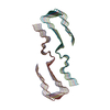
|
|---|---|
| 1 |
|
-Components
| #1: Protein/peptide | Mass: 4335.852 Da / Num. of mol.: 12 Source method: isolated from a genetically manipulated source Source: (gene. exp.) Homo sapiens (human) / Production host:  |
|---|
-Experimental details
-Experiment
| Experiment | Method: ELECTRON MICROSCOPY |
|---|---|
| EM experiment | Aggregation state: HELICAL ARRAY / 3D reconstruction method: helical reconstruction |
- Sample preparation
| Component | Name: seeded in vitro Abeta(1-40) amyloid fibril / Type: COMPLEX / Details: morphology I / Entity ID: all / Source: RECOMBINANT |
|---|---|
| Source (natural) | Organism: Homo sapiens (human) |
| Source (recombinant) | Organism: |
| Buffer solution | pH: 7.4 |
| Buffer component | Conc.: 100 mM / Name: Disodium phosphate / Formula: Na2HPO4 |
| Specimen | Conc.: 0.005 mg/ml / Embedding applied: NO / Shadowing applied: NO / Staining applied: NO / Vitrification applied: YES |
| Specimen support | Grid material: COPPER / Grid mesh size: 400 divisions/in. / Grid type: C-flat-1.2/1.3 |
| Vitrification | Instrument: LEICA PLUNGER / Cryogen name: ETHANE / Humidity: 95 % |
- Electron microscopy imaging
| Experimental equipment |  Model: Titan Krios / Image courtesy: FEI Company |
|---|---|
| Microscopy | Model: FEI TITAN KRIOS |
| Electron gun | Electron source:  FIELD EMISSION GUN / Accelerating voltage: 300 kV / Illumination mode: FLOOD BEAM FIELD EMISSION GUN / Accelerating voltage: 300 kV / Illumination mode: FLOOD BEAM |
| Electron lens | Mode: BRIGHT FIELD / Nominal magnification: 130000 X / Nominal defocus max: 2000 nm / Nominal defocus min: 800 nm / Cs: 2.7 mm |
| Specimen holder | Cryogen: NITROGEN |
| Image recording | Average exposure time: 10 sec. / Electron dose: 53.79 e/Å2 / Detector mode: COUNTING / Film or detector model: GATAN K2 QUANTUM (4k x 4k) / Num. of grids imaged: 1 / Num. of real images: 3018 |
- Processing
| EM software |
| ||||||||||||||||||||||||||||
|---|---|---|---|---|---|---|---|---|---|---|---|---|---|---|---|---|---|---|---|---|---|---|---|---|---|---|---|---|---|
| CTF correction | Type: PHASE FLIPPING AND AMPLITUDE CORRECTION | ||||||||||||||||||||||||||||
| Helical symmerty | Angular rotation/subunit: -178.82 ° / Axial rise/subunit: 2.38 Å / Axial symmetry: C1 | ||||||||||||||||||||||||||||
| Particle selection | Num. of particles selected: 207732 | ||||||||||||||||||||||||||||
| 3D reconstruction | Resolution: 2.97 Å / Resolution method: FSC 0.143 CUT-OFF / Num. of particles: 114218 / Symmetry type: HELICAL | ||||||||||||||||||||||||||||
| Atomic model building | Protocol: BACKBONE TRACE / Space: REAL / Target criteria: corrleation coefficient | ||||||||||||||||||||||||||||
| Atomic model building | PDB-ID: 6SHS Accession code: 6SHS / Source name: PDB / Type: experimental model |

