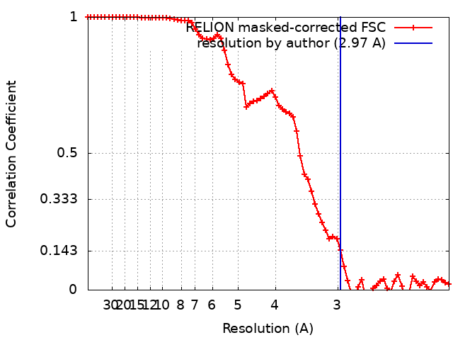
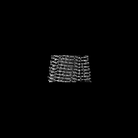
Journal: J Mol Biol / Year: 2024 Title: Cryo-EM Analysis of the Effect of Seeding with Brain-derived Aβ Amyloid Fibrils. Authors: Peter Benedikt Pfeiffer / Marijana Ugrina / Nadine Schwierz / Christina J Sigurdson / Matthias Schmidt / Marcus Fändrich / Abstract: Aβ amyloid fibrils from Alzheimer's brain tissue are polymorphic and structurally different from typical in vitro formed Aβ fibrils. Here, we show that brain-derived (ex vivo) fibril structures can ...Aβ amyloid fibrils from Alzheimer's brain tissue are polymorphic and structurally different from typical in vitro formed Aβ fibrils. Here, we show that brain-derived (ex vivo) fibril structures can be proliferated by seeding in vitro. The proliferation reaction is only efficient for one of the three abundant ex vivo Aβ fibril morphologies, which consists of two peptide stacks, while the inefficiently proliferated fibril morphologies contain four or six peptide stacks. In addition to the seeded fibril structures, we find that de novo nucleated fibril structures can emerge in seeded samples if the seeding reaction is continued over multiple generations. These data imply a competition between de novo nucleation and seed extension and suggest further that seeding favours the outgrowth of fibril morphologies that contain fewer peptide stacks.
Film or detector model: GATAN K2 QUANTUM (4k x 4k) / Detector mode: COUNTING / Number grids imaged: 1 / Number real images: 3018 / Average exposure time: 10.0 sec. / Average electron dose: 53.79 e/Å2
Electron beam
Acceleration voltage: 300 kV / Electron source: FIELD EMISSION GUN
In the structure databanks used in Yorodumi, some data are registered as the other names, "COVID-19 virus" and "2019-nCoV". Here are the details of the virus and the list of structure data.
Jan 31, 2019. EMDB accession codes are about to change! (news from PDBe EMDB page)
EMDB accession codes are about to change! (news from PDBe EMDB page)
The allocation of 4 digits for EMDB accession codes will soon come to an end. Whilst these codes will remain in use, new EMDB accession codes will include an additional digit and will expand incrementally as the available range of codes is exhausted. The current 4-digit format prefixed with “EMD-” (i.e. EMD-XXXX) will advance to a 5-digit format (i.e. EMD-XXXXX), and so on. It is currently estimated that the 4-digit codes will be depleted around Spring 2019, at which point the 5-digit format will come into force.
The EM Navigator/Yorodumi systems omit the EMD- prefix.
Related info.:Q: What is EMD? / ID/Accession-code notation in Yorodumi/EM Navigator
Yorodumi is a browser for structure data from EMDB, PDB, SASBDB, etc.
This page is also the successor to EM Navigator detail page, and also detail information page/front-end page for Omokage search.
The word "yorodu" (or yorozu) is an old Japanese word meaning "ten thousand". "mi" (miru) is to see.
Related info.:EMDB / PDB / SASBDB / Comparison of 3 databanks / Yorodumi Search / Aug 31, 2016. New EM Navigator & Yorodumi / Yorodumi Papers / Jmol/JSmol / Function and homology information / Changes in new EM Navigator and Yorodumi
 Movie
Movie Controller


 Open data
 Basic information
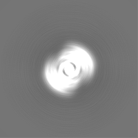
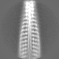
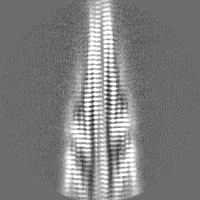
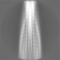
 Map data
 Sample
 Keywords
 Function and homology information
 Homo sapiens (human)
 Authors
 Germany, 1 items
  Citation
 Structure visualization
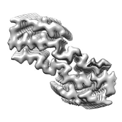
 Downloads & links
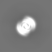
 emd_17168.png
 http://ftp.pdbj.org/pub/emdb/structures/EMD-17168


 Z (Sec.)
 Y (Row.)
 X (Col.)


 Sample components
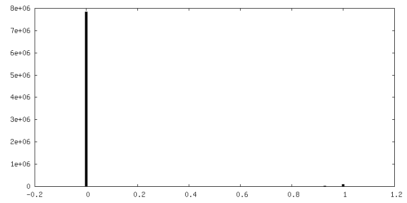
 Processing
 Electron microscopy
 FIELD EMISSION GUN