 Movie
Movie Controller
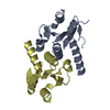

[English] 日本語
 Yorodumi
- PDB-8osc: Structure of Homo sapiens 2'-deoxynucleoside 5'-phosphate N-hydro... -
+ Open data

- Basic information
| Entry | Database: PDB / ID: 8osc | ||||||
|---|---|---|---|---|---|---|---|
| Title | Structure of Homo sapiens 2'-deoxynucleoside 5'-phosphate N-hydrolase 1 (DNPH1) bound to deoxyuridine 5'- monophosphate | ||||||
 Components Components | 2'-deoxynucleoside 5'-phosphate N-hydrolase 1 | ||||||
 Keywords Keywords | HYDROLASE / deoxyribonucleoside 5'-monophosphate N-glycosidase activity | ||||||
| Function / homology |  Function and homology information Function and homology informationpurine nucleotide catabolic process / 5-hydroxymethyl-dUMP N-hydrolase activity / nucleoside salvage / deoxyribonucleoside monophosphate catabolic process / dGMP catabolic process / Purine catabolism / allantoin metabolic process / Hydrolases; Glycosylases; Hydrolysing N-glycosyl compounds / epithelial cell differentiation / positive regulation of cell growth ...purine nucleotide catabolic process / 5-hydroxymethyl-dUMP N-hydrolase activity / nucleoside salvage / deoxyribonucleoside monophosphate catabolic process / dGMP catabolic process / Purine catabolism / allantoin metabolic process / Hydrolases; Glycosylases; Hydrolysing N-glycosyl compounds / epithelial cell differentiation / positive regulation of cell growth / protein homodimerization activity / extracellular exosome / identical protein binding / nucleus / cytoplasm / cytosol Similarity search - Function | ||||||
| Biological species |  Homo sapiens (human) Homo sapiens (human) | ||||||
| Method |  X-RAY DIFFRACTION / SYNCHROTRON / MOLECULAR REPLACEMENT / Resolution: 1.42 Å X-RAY DIFFRACTION / SYNCHROTRON / MOLECULAR REPLACEMENT / Resolution: 1.42 Å | ||||||
 Authors Authors | Devi, S. / da Silva, R.G. | ||||||
| Funding support |  United Kingdom, 1items United Kingdom, 1items
| ||||||
 Citation Citation | Journal: Biochemistry / Year: 2023 Title: Human 2'-Deoxynucleoside 5'-Phosphate N -Hydrolase 1: Mechanism of 2'-Deoxyuridine 5'-Monophosphate Hydrolysis. Authors: Devi, S. / Carberry, A.E. / Zickuhr, G.M. / Dickson, A.L. / Harrison, D.J. / da Silva, R.G. | ||||||
| History |
|
- Structure visualization
| Structure viewer | Molecule: MolmilJmol/JSmol |
|---|
- Downloads & links
-Download
| PDBx/mmCIF format | 8osc.cif.gz | 69.1 KB | Display | PDBx/mmCIF format |
|---|---|---|---|---|
| PDB format | pdb8osc.ent.gz | 47.9 KB | Display | PDB format |
| PDBx/mmJSON format | 8osc.json.gz | Tree view | PDBx/mmJSON format | |
| Others |  Other downloads Other downloads |
-Validation report
| Arichive directory | https://data.pdbj.org/pub/pdb/validation_reports/os/8oscftp://data.pdbj.org/pub/pdb/validation_reports/os/8osc | HTTPS FTP |
|---|
-Related structure data
-Links
 PDBj
PDBj- Assembly
| Deposited unit | 
| |||||||||
|---|---|---|---|---|---|---|---|---|---|---|
| 1 |
| |||||||||
| Unit cell |
| |||||||||
| Components on special symmetry positions |
|
-Components
| #1: Protein | Mass: 16194.220 Da / Num. of mol.: 2 Source method: isolated from a genetically manipulated source Source: (gene. exp.) Homo sapiens (human) / Gene: DNPH1, C6orf108, RCL / Production host:  References: UniProt: O43598, Hydrolases; Glycosylases; Hydrolysing N-glycosyl compounds #2: Chemical | ChemComp-UMP / |   Mass: 308.182 Da / Num. of mol.: 1 / Source method: obtained synthetically / Formula: C9H13N2O8P / Feature type: SUBJECT OF INVESTIGATION Mass: 308.182 Da / Num. of mol.: 1 / Source method: obtained synthetically / Formula: C9H13N2O8P / Feature type: SUBJECT OF INVESTIGATION#3: Water | ChemComp-HOH / |  Mass: 18.015 Da / Num. of mol.: 192 / Source method: isolated from a natural source / Formula: H2O Mass: 18.015 Da / Num. of mol.: 192 / Source method: isolated from a natural source / Formula: H2OHas ligand of interest | Y | |
|---|
-Experimental details
-Experiment
| Experiment | Method: X-RAY DIFFRACTION / Number of used crystals: 1 |
|---|
- Sample preparation
| Crystal | Density Matthews: 1.84 Å3/Da / Density % sol: 33.04 % |
|---|---|
| Crystal grow | Temperature: 293.15 K / Method: vapor diffusion, hanging drop / pH: 4 Details: [40 mM sodium propionate, 20 mM MES and 40 mM BIS-TRIS propane] pH 4.0 and 20% PEG 1500 |
-Data collection
| Diffraction | Mean temperature: 100 K / Serial crystal experiment: N |
|---|---|
| Diffraction source | Source: SYNCHROTRON / Site: Diamond / Beamline: I04 / Wavelength: 0.9537 Å |
| Detector | Type: DECTRIS EIGER2 XE 16M / Detector: PIXEL / Date: Oct 8, 2022 |
| Radiation | Monochromator: M / Protocol: SINGLE WAVELENGTH / Monochromatic (M) / Laue (L): M / Scattering type: x-ray |
| Radiation wavelength | Wavelength: 0.9537 Å / Relative weight: 1 |
| Reflection | Resolution: 1.42→33.38 Å / Num. obs: 44559 / % possible obs: 99.7 % / Redundancy: 15.4 % / Biso Wilson estimate: 19.39 Å2 / CC1/2: 0.999 / Rmerge(I) obs: 0.058 / Rpim(I) all: 0.015 / Rrim(I) all: 0.06 / Net I/σ(I): 24.7 |
| Reflection shell | Resolution: 1.42→1.44 Å / Redundancy: 10.9 % / Rmerge(I) obs: 0.814 / Mean I/σ(I) obs: 4 / Num. unique obs: 2097 / CC1/2: 0.899 / Rpim(I) all: 0.254 / Rrim(I) all: 0.855 / % possible all: 94.9 |
- Processing
| Software |
| |||||||||||||||||||||||||||||||||||||||||||||||||||||||||||||||||||||||||||||||||||||||||||||||||||||||||||||||||||||||
|---|---|---|---|---|---|---|---|---|---|---|---|---|---|---|---|---|---|---|---|---|---|---|---|---|---|---|---|---|---|---|---|---|---|---|---|---|---|---|---|---|---|---|---|---|---|---|---|---|---|---|---|---|---|---|---|---|---|---|---|---|---|---|---|---|---|---|---|---|---|---|---|---|---|---|---|---|---|---|---|---|---|---|---|---|---|---|---|---|---|---|---|---|---|---|---|---|---|---|---|---|---|---|---|---|---|---|---|---|---|---|---|---|---|---|---|---|---|---|---|---|
| Refinement | Method to determine structure: MOLECULAR REPLACEMENT / Resolution: 1.42→33.38 Å / SU ML: 0.15 / Cross valid method: FREE R-VALUE / σ(F): 1.41 / Phase error: 21.65 / Stereochemistry target values: ML
| |||||||||||||||||||||||||||||||||||||||||||||||||||||||||||||||||||||||||||||||||||||||||||||||||||||||||||||||||||||||
| Solvent computation | Shrinkage radii: 0.9 Å / VDW probe radii: 1.1 Å / Solvent model: FLAT BULK SOLVENT MODEL | |||||||||||||||||||||||||||||||||||||||||||||||||||||||||||||||||||||||||||||||||||||||||||||||||||||||||||||||||||||||
| Refinement step | Cycle: LAST / Resolution: 1.42→33.38 Å
| |||||||||||||||||||||||||||||||||||||||||||||||||||||||||||||||||||||||||||||||||||||||||||||||||||||||||||||||||||||||
| Refine LS restraints |
| |||||||||||||||||||||||||||||||||||||||||||||||||||||||||||||||||||||||||||||||||||||||||||||||||||||||||||||||||||||||
| LS refinement shell |
|
