- PDB-8or1: Co-crystal strucutre of PD-L1 with low molecular weight inhibitor -
+
Open data
ID or keywords:
Loading...
-
Basic information
Entry
Database: PDB / ID: 8or1
Title
Co-crystal strucutre of PD-L1 with low molecular weight inhibitor
Components
Programmed cell death 1 ligand 1
Keywords
IMMUNE SYSTEM / PD-L1 / inhibitor / immunotheraphy
Function / homology
Function and homology information
negative regulation of tumor necrosis factor superfamily cytokine production / positive regulation of activated CD8-positive, alpha-beta T cell apoptotic process / negative regulation of CD8-positive, alpha-beta T cell activation / negative regulation of T cell mediated immune response to tumor cell / TRIF-dependent toll-like receptor signaling pathway / negative regulation of CD4-positive, alpha-beta T cell proliferation / STAT3 nuclear events downstream of ALK signaling / negative regulation of interleukin-10 production / negative regulation of activated T cell proliferation / negative regulation of type II interferon production ...negative regulation of tumor necrosis factor superfamily cytokine production / positive regulation of activated CD8-positive, alpha-beta T cell apoptotic process / negative regulation of CD8-positive, alpha-beta T cell activation / negative regulation of T cell mediated immune response to tumor cell / TRIF-dependent toll-like receptor signaling pathway / negative regulation of CD4-positive, alpha-beta T cell proliferation / STAT3 nuclear events downstream of ALK signaling / negative regulation of interleukin-10 production / negative regulation of activated T cell proliferation / negative regulation of type II interferon production / positive regulation of interleukin-10 production / Co-inhibition by PD-1 / negative regulation of T cell proliferation / T cell costimulation / response to cytokine / positive regulation of T cell proliferation / recycling endosome membrane / actin cytoskeleton / cellular response to lipopolysaccharide / early endosome membrane / adaptive immune response / transcription coactivator activity / cell surface receptor signaling pathway / immune response / receptor ligand activity / external side of plasma membrane / signal transduction / extracellular exosome / nucleoplasm / plasma membrane Similarity search - Function
Method to determine structure: MOLECULAR REPLACEMENT / Resolution: 3.5→38.13 Å / Cor.coef. Fo:Fc: 0.927 / Cor.coef. Fo:Fc free: 0.906 / SU B: 122.631 / SU ML: 0.765 / Cross valid method: THROUGHOUT / ESU R Free: 0.781 / Stereochemistry target values: MAXIMUM LIKELIHOOD Details: U VALUES : WITH TLS ADDED HYDROGENS HAVE BEEN ADDED IN THE RIDING POSITIONS U VALUES : RESIDUAL ONLY
Rfactor
Num. reflection
% reflection
Selection details
Rfree
0.29624
385
9.7 %
RANDOM
Rwork
0.29228
-
-
-
obs
0.29265
3590
99.25 %
-
Solvent computation
Ion probe radii: 0.9 Å / Shrinkage radii: 0.9 Å / VDW probe radii: 1.2 Å / Solvent model: MASK
Displacement parameters
Biso mean: 99.888 Å2
Baniso -1
Baniso -2
Baniso -3
1-
-1.16 Å2
-0.58 Å2
-0 Å2
2-
-
-1.16 Å2
0 Å2
3-
-
-
3.77 Å2
Refinement step
Cycle: LAST / Resolution: 3.5→38.13 Å
Protein
Nucleic acid
Ligand
Solvent
Total
Num. atoms
1858
0
37
0
1895
Refine LS restraints
Refine-ID
Type
Dev ideal
Dev ideal target
Number
X-RAY DIFFRACTION
r_bond_refined_d
0.006
0.017
1930
X-RAY DIFFRACTION
r_bond_other_d
0.001
0.016
1797
X-RAY DIFFRACTION
r_angle_refined_deg
0.937
1.821
2609
X-RAY DIFFRACTION
r_angle_other_deg
0.328
1.565
4180
X-RAY DIFFRACTION
r_dihedral_angle_1_deg
7.287
5.208
240
X-RAY DIFFRACTION
r_dihedral_angle_3_deg
13.365
10
346
X-RAY DIFFRACTION
r_chiral_restr
0.264
0.2
290
X-RAY DIFFRACTION
r_gen_planes_refined
0.003
0.02
2154
X-RAY DIFFRACTION
r_gen_planes_other
0.001
0.02
373
LS refinement shell
Resolution: 3.501→3.591 Å / Total num. of bins used: 20
Rfactor
Num. reflection
% reflection
Rfree
0.435
22
-
Rwork
0.424
258
-
obs
-
-
100 %
Refinement TLS params.
Method: refined / Refine-ID: X-RAY DIFFRACTION
ID
L11 (°2)
L12 (°2)
L13 (°2)
L22 (°2)
L23 (°2)
L33 (°2)
S11 (Å °)
S12 (Å °)
S13 (Å °)
S21 (Å °)
S22 (Å °)
S23 (Å °)
S31 (Å °)
S32 (Å °)
S33 (Å °)
T11 (Å2)
T12 (Å2)
T13 (Å2)
T22 (Å2)
T23 (Å2)
T33 (Å2)
Origin x (Å)
Origin y (Å)
Origin z (Å)
1
10.6365
4.9011
-1.3764
5.2904
-0.7922
2.1025
0.3262
-0.7768
-0.3007
0.2815
-0.279
-0.9065
-0.0448
0.0033
-0.0472
0.4244
0.1703
0.0306
0.4704
0.1556
0.2648
-35.148
27.604
-9.749
2
10.7473
-1.1878
-0.4445
5.8732
-2.0858
4.3447
0.0365
-0.55
1.2377
0.3724
0.1247
0.709
-0.8914
-1.0118
-0.1612
0.4755
0.2883
0.2107
0.3361
-0.0735
0.5354
-38.12
50.487
-20.798
Refinement TLS group
ID
Refine-ID
Refine TLS-ID
Auth asym-ID
Auth seq-ID
1
X-RAY DIFFRACTION
1
A
4 - 119
2
X-RAY DIFFRACTION
2
B
4 - 119
+
About Yorodumi
-
News
-
Feb 9, 2022. New format data for meta-information of EMDB entries
New format data for meta-information of EMDB entries
Version 3 of the EMDB header file is now the official format.
The previous official version 1.9 will be removed from the archive.
In the structure databanks used in Yorodumi, some data are registered as the other names, "COVID-19 virus" and "2019-nCoV". Here are the details of the virus and the list of structure data.
Jan 31, 2019. EMDB accession codes are about to change! (news from PDBe EMDB page)
EMDB accession codes are about to change! (news from PDBe EMDB page)
The allocation of 4 digits for EMDB accession codes will soon come to an end. Whilst these codes will remain in use, new EMDB accession codes will include an additional digit and will expand incrementally as the available range of codes is exhausted. The current 4-digit format prefixed with “EMD-” (i.e. EMD-XXXX) will advance to a 5-digit format (i.e. EMD-XXXXX), and so on. It is currently estimated that the 4-digit codes will be depleted around Spring 2019, at which point the 5-digit format will come into force.
The EM Navigator/Yorodumi systems omit the EMD- prefix.
Related info.:Q: What is EMD? / ID/Accession-code notation in Yorodumi/EM Navigator
Yorodumi is a browser for structure data from EMDB, PDB, SASBDB, etc.
This page is also the successor to EM Navigator detail page, and also detail information page/front-end page for Omokage search.
The word "yorodu" (or yorozu) is an old Japanese word meaning "ten thousand". "mi" (miru) is to see.
Related info.:EMDB / PDB / SASBDB / Comparison of 3 databanks / Yorodumi Search / Aug 31, 2016. New EM Navigator & Yorodumi / Yorodumi Papers / Jmol/JSmol / Function and homology information / Changes in new EM Navigator and Yorodumi
 Movie
Movie Controller

 Yorodumi
 Open data
 Basic information
 Components
 Keywords
 Function and homology information
 Homo sapiens (human)
 X-RAY DIFFRACTION /
  Authors
 China,
  Poland, 3items
  Citation
 Structure visualization
 Downloads & links
 Other downloads
 PDBj
PDBj


 Assembly

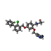
 Mass: 517.979 Da / Num. of mol.: 1 / Source method: obtained synthetically / Formula: C29H25ClFN3O3 / Feature type: SUBJECT OF INVESTIGATION
 Sample preparation
 / Beamline: P11 / Wavelength: 1.03321 Å
 Processing