 Movie
Movie Controller

[English] 日本語
 Yorodumi
- PDB-8oog: Crystal structure of human MAT2a with S-Adenosylmethionine and a ... -
+ Open data

- Basic information
| Entry | Database: PDB / ID: 8oog | ||||||
|---|---|---|---|---|---|---|---|
| Title | Crystal structure of human MAT2a with S-Adenosylmethionine and a fragment bound in a novel pocket | ||||||
 Components Components | S-adenosylmethionine synthase isoform type-2 | ||||||
 Keywords Keywords | TRANSFERASE / methionine adenosyltransferase / s-adenosylmethionine synthetase / fragment screen / allosteric binder | ||||||
| Function / homology |  Function and homology information Function and homology informationmethionine adenosyltransferase complex / methionine adenosyltransferase / methionine adenosyltransferase activity / S-adenosylmethionine biosynthetic process / protein heterooligomerization / Methylation / cellular response to methionine / protein hexamerization / small molecule binding / one-carbon metabolic process ...methionine adenosyltransferase complex / methionine adenosyltransferase / methionine adenosyltransferase activity / S-adenosylmethionine biosynthetic process / protein heterooligomerization / Methylation / cellular response to methionine / protein hexamerization / small molecule binding / one-carbon metabolic process / positive regulation of TORC1 signaling / cellular response to leukemia inhibitory factor / ATP binding / metal ion binding / identical protein binding / cytosol Similarity search - Function | ||||||
| Biological species |  Homo sapiens (human) Homo sapiens (human) | ||||||
| Method |  X-RAY DIFFRACTION / SYNCHROTRON / MOLECULAR REPLACEMENT / Resolution: 1.384 Å X-RAY DIFFRACTION / SYNCHROTRON / MOLECULAR REPLACEMENT / Resolution: 1.384 Å | ||||||
 Authors Authors | Schimpl, M. | ||||||
| Funding support | 1items
| ||||||
 Citation Citation | Journal: Chem Sci / Year: 2023 Title: Combining structural and coevolution information to unveil allosteric sites. Authors: La Sala, G. / Pfleger, C. / Kack, H. / Wissler, L. / Nevin, P. / Bohm, K. / Janet, J.P. / Schimpl, M. / Stubbs, C.J. / De Vivo, M. / Tyrchan, C. / Hogner, A. / Gohlke, H. / Frolov, A.I. | ||||||
| History |
|
- Structure visualization
| Structure viewer | Molecule: MolmilJmol/JSmol |
|---|
- Downloads & links
-Download
| PDBx/mmCIF format | 8oog.cif.gz | 159 KB | Display | PDBx/mmCIF format |
|---|---|---|---|---|
| PDB format | pdb8oog.ent.gz | 123.2 KB | Display | PDB format |
| PDBx/mmJSON format | 8oog.json.gz | Tree view | PDBx/mmJSON format | |
| Others |  Other downloads Other downloads |
-Validation report
| Summary document | 8oog_validation.pdf.gz | 974.7 KB | Display | wwPDB validaton report |
|---|---|---|---|---|
| Full document | 8oog_full_validation.pdf.gz | 974.6 KB | Display | |
| Data in XML | 8oog_validation.xml.gz | 16.3 KB | Display | |
| Data in CIF | 8oog_validation.cif.gz | 23.8 KB | Display | |
| Arichive directory | https://data.pdbj.org/pub/pdb/validation_reports/oo/8oogftp://data.pdbj.org/pub/pdb/validation_reports/oo/8oog | HTTPS FTP |
-Related structure data
| Similar structure data |
|---|
-Links
 PDBj
PDBj
- Assembly
| Deposited unit | 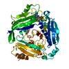
| |||||||||
|---|---|---|---|---|---|---|---|---|---|---|
| 1 |
| |||||||||
| Unit cell |
| |||||||||
| Components on special symmetry positions |
|
-Components
| #1: Protein | Mass: 43935.828 Da / Num. of mol.: 1 / Fragment: full length protein Source method: isolated from a genetically manipulated source Source: (gene. exp.) Homo sapiens (human) / Gene: MAT2A, AMS2, MATA2 / Production host:  | ||||
|---|---|---|---|---|---|
| #2: Chemical | ChemComp-SAM /   Mass: 398.437 Da / Num. of mol.: 1 / Source method: obtained synthetically / Formula: C15H22N6O5S Mass: 398.437 Da / Num. of mol.: 1 / Source method: obtained synthetically / Formula: C15H22N6O5S | ||||
| #3: Chemical | ChemComp-VUO / 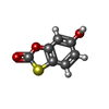  Mass: 168.170 Da / Num. of mol.: 1 / Source method: obtained synthetically / Formula: C7H4O3S / Feature type: SUBJECT OF INVESTIGATION Mass: 168.170 Da / Num. of mol.: 1 / Source method: obtained synthetically / Formula: C7H4O3S / Feature type: SUBJECT OF INVESTIGATION | ||||
| #4: Chemical |   Mass: 78.133 Da / Num. of mol.: 2 / Source method: obtained synthetically / Formula: C2H6OS / Comment: DMSO, precipitant*YM Mass: 78.133 Da / Num. of mol.: 2 / Source method: obtained synthetically / Formula: C2H6OS / Comment: DMSO, precipitant*YM#5: Water | ChemComp-HOH / |  Mass: 18.015 Da / Num. of mol.: 176 / Source method: isolated from a natural source / Formula: H2O Mass: 18.015 Da / Num. of mol.: 176 / Source method: isolated from a natural source / Formula: H2OHas ligand of interest | Y | |
-Experimental details
-Experiment
| Experiment | Method: X-RAY DIFFRACTION / Number of used crystals: 1 |
|---|
- Sample preparation
| Crystal | Density Matthews: 2.13 Å3/Da / Density % sol: 42.2 % |
|---|---|
| Crystal grow | Temperature: 293 K / Method: vapor diffusion / pH: 8 Details: 8 % PEG8000, 12 % ethylene glycol, 0.1 M Hepes pH 8.0 |
-Data collection
| Diffraction | Mean temperature: 100 K / Serial crystal experiment: N |
|---|---|
| Diffraction source | Source: SYNCHROTRON / Site: SOLEIL  / Beamline: PROXIMA 2 / Wavelength: 0.98011 Å / Beamline: PROXIMA 2 / Wavelength: 0.98011 Å |
| Detector | Type: DECTRIS EIGER X 9M / Detector: PIXEL / Date: Mar 30, 2023 |
| Radiation | Protocol: SINGLE WAVELENGTH / Monochromatic (M) / Laue (L): M / Scattering type: x-ray |
| Radiation wavelength | Wavelength: 0.98011 Å / Relative weight: 1 |
| Reflection | Resolution: 1.384→73.374 Å / Num. obs: 75517 / % possible obs: 98.7 % / Redundancy: 6.2 % / Rmerge(I) obs: 0.07 / Rpim(I) all: 0.031 / Rrim(I) all: 0.077 / Net I/σ(I): 13.3 / Num. measured all: 466961 |
| Reflection shell | Resolution: 1.384→1.408 Å / % possible obs: 97.2 % / Redundancy: 6.4 % / Rmerge(I) obs: 1.097 / Num. measured all: 23485 / Num. unique obs: 3671 / Rpim(I) all: 0.466 / Rrim(I) all: 1.194 / Net I/σ(I) obs: 2.1 |
- Processing
| Software |
| ||||||||||||||||||||||||||||||||||||||||||||||||||||||||||||||||||||||||||||||||||||||||||||||||||||||||||||||||||
|---|---|---|---|---|---|---|---|---|---|---|---|---|---|---|---|---|---|---|---|---|---|---|---|---|---|---|---|---|---|---|---|---|---|---|---|---|---|---|---|---|---|---|---|---|---|---|---|---|---|---|---|---|---|---|---|---|---|---|---|---|---|---|---|---|---|---|---|---|---|---|---|---|---|---|---|---|---|---|---|---|---|---|---|---|---|---|---|---|---|---|---|---|---|---|---|---|---|---|---|---|---|---|---|---|---|---|---|---|---|---|---|---|---|---|---|
| Refinement | Method to determine structure: MOLECULAR REPLACEMENT / Resolution: 1.384→73.37 Å / Cor.coef. Fo:Fc: 0.893 / Cor.coef. Fo:Fc free: 0.949 / SU R Cruickshank DPI: 0.059 / Cross valid method: THROUGHOUT / σ(F): 0 / SU R Blow DPI: 0.059 / SU Rfree Blow DPI: 0.057 / SU Rfree Cruickshank DPI: 0.057
| ||||||||||||||||||||||||||||||||||||||||||||||||||||||||||||||||||||||||||||||||||||||||||||||||||||||||||||||||||
| Displacement parameters | Biso mean: 19.35 Å2
| ||||||||||||||||||||||||||||||||||||||||||||||||||||||||||||||||||||||||||||||||||||||||||||||||||||||||||||||||||
| Refine analyze | Luzzati coordinate error obs: 0.18 Å | ||||||||||||||||||||||||||||||||||||||||||||||||||||||||||||||||||||||||||||||||||||||||||||||||||||||||||||||||||
| Refinement step | Cycle: LAST / Resolution: 1.384→73.37 Å
| ||||||||||||||||||||||||||||||||||||||||||||||||||||||||||||||||||||||||||||||||||||||||||||||||||||||||||||||||||
| Refine LS restraints |
| ||||||||||||||||||||||||||||||||||||||||||||||||||||||||||||||||||||||||||||||||||||||||||||||||||||||||||||||||||
| LS refinement shell | Resolution: 1.384→1.39 Å
| ||||||||||||||||||||||||||||||||||||||||||||||||||||||||||||||||||||||||||||||||||||||||||||||||||||||||||||||||||
| Refinement TLS params. | Method: refined / Origin x: 26.0243 Å / Origin y: 38.907 Å / Origin z: 32.6625 Å
| ||||||||||||||||||||||||||||||||||||||||||||||||||||||||||||||||||||||||||||||||||||||||||||||||||||||||||||||||||
| Refinement TLS group | Selection details: { A|* } |
