| Entry | Database: PDB / ID: 8onj
|
|---|
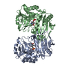
| Title | Crystal structure of D-amino acid aminotransferase from Aminobacterium colombiense point mutant R88L |
|---|
 Components Components | Aminotransferase class IV |
|---|
 Keywords Keywords | TRANSFERASE / DAAT / Transaminase / point mutant / aminotransferase |
|---|
| Function / homology |  Function and homology information Function and homology information
: / Aminotransferase class 4, branched-chain amino acid transferase, N-terminal domain / D-amino Acid Aminotransferase; Chain A, domain 2 / D-amino Acid Aminotransferase, subunit A, domain 2 / Branched-chain-amino-acid aminotransferase-like, N-terminal / Aminotransferase class IV / Aminotransferase-like, PLP-dependent enzymes / Branched-chain-amino-acid aminotransferase-like, C-terminal / Amino-transferase class IV / D-amino Acid Aminotransferase; Chain A, domain 1 ...: / Aminotransferase class 4, branched-chain amino acid transferase, N-terminal domain / D-amino Acid Aminotransferase; Chain A, domain 2 / D-amino Acid Aminotransferase, subunit A, domain 2 / Branched-chain-amino-acid aminotransferase-like, N-terminal / Aminotransferase class IV / Aminotransferase-like, PLP-dependent enzymes / Branched-chain-amino-acid aminotransferase-like, C-terminal / Amino-transferase class IV / D-amino Acid Aminotransferase; Chain A, domain 1 / Alpha-Beta Barrel / 2-Layer Sandwich / Alpha BetaSimilarity search - Domain/homology |
|---|
| Biological species |  Aminobacterium colombiense (bacteria) Aminobacterium colombiense (bacteria) |
|---|
| Method |  X-RAY DIFFRACTION / MOLECULAR REPLACEMENT / Resolution: 1.8 Å X-RAY DIFFRACTION / MOLECULAR REPLACEMENT / Resolution: 1.8 Å |
|---|
 Authors Authors | Matyuta, I.O. / Boyko, K.M. / Minyaev, M.E. / Shilova, S.A. / Bezsudnova, E.Y. / Popov, V.O. |
|---|
| Funding support |  Russian Federation, 1items Russian Federation, 1items | Organization | Grant number | Country |
|---|
| Russian Science Foundation | 19-14-00164 | Russian Federation |
|
|---|
 Citation Citation | Journal: Biochem.J. / Year: 2023
Title: In search for structural targets for engineering d-amino acid transaminase: modulation of pH optimum and substrate specificity.
Authors: Shilova, S.A. / Matyuta, I.O. / Khrenova, M.G. / Nikolaeva, A.Y. / Klyachko, N.L. / Minyaev, M.E. / Khomutov, A.R. / Boyko, K.M. / Popov, V.O. / Bezsudnova, E.Y. |
|---|
| History | | Deposition | Apr 3, 2023 | Deposition site: PDBE / Processing site: PDBE |
|---|
| Revision 1.0 | Aug 30, 2023 | Provider: repository / Type: Initial release |
|---|
|
|---|
 Movie
Movie Controller

 Yorodumi
 Open data
 Basic information
 Structure visualization
 Downloads & links
 Other downloads
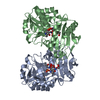

 PDBj
PDBj
 Assembly
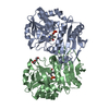

 Mass: 247.142 Da / Num. of mol.: 2 / Source method: obtained synthetically / Formula: C8H10NO6P / Feature type: SUBJECT OF INVESTIGATION
 Mass: 106.120 Da / Num. of mol.: 1 / Source method: obtained synthetically / Formula: C4H10O3
 Mass: 18.015 Da / Num. of mol.: 255 / Source method: isolated from a natural source / Formula: H2O
 Sample preparation
 Processing