Movie
Movie Controller


+ Open data

- Basic information
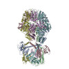
| Entry | Database: PDB / ID: 8ojl | ||||||||||||||||||||||||
|---|---|---|---|---|---|---|---|---|---|---|---|---|---|---|---|---|---|---|---|---|---|---|---|---|---|
| Title | Human Mitochondrial Lon Y394E Mutant ADP Bound | ||||||||||||||||||||||||
 Components Components | Lon protease homolog, mitochondrial | ||||||||||||||||||||||||
 Keywords Keywords | HYDROLASE / Human mitochondrial AAA+ protease / motor protein | ||||||||||||||||||||||||
| Function / homology |  Function and homology information Function and homology informationoxidation-dependent protein catabolic process / response to aluminum ion / PH domain binding / endopeptidase La / mitochondrial protein catabolic process / G-quadruplex DNA binding / ATP-dependent peptidase activity / protein quality control for misfolded or incompletely synthesized proteins / mitochondrial nucleoid / insulin receptor substrate binding ...oxidation-dependent protein catabolic process / response to aluminum ion / PH domain binding / endopeptidase La / mitochondrial protein catabolic process / G-quadruplex DNA binding / ATP-dependent peptidase activity / protein quality control for misfolded or incompletely synthesized proteins / mitochondrial nucleoid / insulin receptor substrate binding / Mitochondrial unfolded protein response (UPRmt) / chaperone-mediated protein complex assembly / DNA polymerase binding / response to hormone / negative regulation of insulin receptor signaling pathway / Mitochondrial protein degradation / : / mitochondrion organization / ADP binding / single-stranded DNA binding / cellular response to oxidative stress / sequence-specific DNA binding / response to hypoxia / single-stranded RNA binding / mitochondrial matrix / serine-type endopeptidase activity / ATP hydrolysis activity / mitochondrion / nucleoplasm / ATP binding / membrane / identical protein binding / cytosol Similarity search - Function | ||||||||||||||||||||||||
| Biological species |  Homo sapiens (human) Homo sapiens (human) | ||||||||||||||||||||||||
| Method | ELECTRON MICROSCOPY / single particle reconstruction / cryo EM / Resolution: 2.88 Å | ||||||||||||||||||||||||
 Authors Authors | Kereiche, S. / Bauer, J.A. / Matyas, P. / Novacek, J. / Kutejova, E. | ||||||||||||||||||||||||
| Funding support | European Union,  Czech Republic, 7items Czech Republic, 7items
| ||||||||||||||||||||||||
 Citation Citation | Journal: Sci Rep / Year: 2024 Title: Polyphosphate and tyrosine phosphorylation in the N-terminal domain of the human mitochondrial Lon protease disrupts its functions. Authors: Nina Kunová / Gabriela Ondrovičová / Jacob A Bauer / Veronika Krajčovičová / Matyáš Pinkas / Barbora Stojkovičová / Henrieta Havalová / Veronika Lukáčová / Lenka Kohútová / ...Authors: Nina Kunová / Gabriela Ondrovičová / Jacob A Bauer / Veronika Krajčovičová / Matyáš Pinkas / Barbora Stojkovičová / Henrieta Havalová / Veronika Lukáčová / Lenka Kohútová / Július Košťan / Lucia Martináková / Peter Baráth / Jiří Nováček / Sebastian Zoll / Sami Kereïche / Eva Kutejová / Vladimír Pevala /   Abstract: Phosphorylation plays a crucial role in the regulation of many fundamental cellular processes. Phosphorylation levels are increased in many cancer cells where they may promote changes in ...Phosphorylation plays a crucial role in the regulation of many fundamental cellular processes. Phosphorylation levels are increased in many cancer cells where they may promote changes in mitochondrial homeostasis. Proteomic studies on various types of cancer identified 17 phosphorylation sites within the human ATP-dependent protease Lon, which degrades misfolded, unassembled and oxidatively damaged proteins in mitochondria. Most of these sites were found in Lon's N-terminal (NTD) and ATPase domains, though little is known about the effects on their function. By combining the biochemical and cryo-electron microscopy studies, we show the effect of Tyr186 and Tyr394 phosphorylations in Lon's NTD, which greatly reduce all Lon activities without affecting its ability to bind substrates or perturbing its tertiary structure. A substantial reduction in Lon's activities is also observed in the presence of polyphosphate, whose amount significantly increases in cancer cells. Our study thus provides an insight into the possible fine-tuning of Lon activities in human diseases, which highlights Lon's importance in maintaining proteostasis in mitochondria. | ||||||||||||||||||||||||
| History |
|
- Structure visualization
| Structure viewer | Molecule: MolmilJmol/JSmol |
|---|
- Downloads & links
-Download
| PDBx/mmCIF format | 8ojl.cif.gz | 2.1 MB | Display | PDBx/mmCIF format |
|---|---|---|---|---|
| PDB format | pdb8ojl.ent.gz | 1.4 MB | Display | PDB format |
| PDBx/mmJSON format | 8ojl.json.gz | Tree view | PDBx/mmJSON format | |
| Others |  Other downloads Other downloads |
-Validation report
| Arichive directory | https://data.pdbj.org/pub/pdb/validation_reports/oj/8ojlftp://data.pdbj.org/pub/pdb/validation_reports/oj/8ojl | HTTPS FTP |
|---|
-Related structure data
| Related structure data |  16915MC 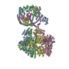 8okaC 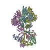 8om7C  8ovfC  8ovgC M: map data used to model this data C: citing same article ( |
|---|---|
| Similar structure data |

-Links
 PDBj
PDBj
- Assembly
| Deposited unit | 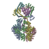
|
|---|---|
| 1 |
|
-Components
| #1: Protein | Mass: 98165.789 Da / Num. of mol.: 6 / Mutation: Y394E Source method: isolated from a genetically manipulated source Source: (gene. exp.) Homo sapiens (human) / Gene: LONP1, PRSS15 / Production host:  #2: Chemical | ChemComp-ADP /   Mass: 427.201 Da / Num. of mol.: 6 / Source method: obtained synthetically / Formula: C10H15N5O10P2 / Comment: ADP, energy-carrying molecule*YM Mass: 427.201 Da / Num. of mol.: 6 / Source method: obtained synthetically / Formula: C10H15N5O10P2 / Comment: ADP, energy-carrying molecule*YMHas ligand of interest | N | |
|---|
-Experimental details
-Experiment
| Experiment | Method: ELECTRON MICROSCOPY |
|---|---|
| EM experiment | Aggregation state: PARTICLE / 3D reconstruction method: single particle reconstruction |
- Sample preparation
| Component | Name: Human mitochondrial Lon protease / Type: COMPLEX / Entity ID: #1 / Source: RECOMBINANT |
|---|---|
| Molecular weight | Experimental value: NO |
| Source (natural) | Organism: Homo sapiens (human) / Organelle: mitochondria |
| Source (recombinant) | Organism: |
| Buffer solution | pH: 7 |
| Specimen | Embedding applied: NO / Shadowing applied: NO / Staining applied: NO / Vitrification applied: YES |
| Vitrification | Cryogen name: ETHANE |
- Electron microscopy imaging
| Experimental equipment |  Model: Titan Krios / Image courtesy: FEI Company |
|---|---|
| Microscopy | Model: FEI TITAN KRIOS |
| Electron gun | Electron source:  FIELD EMISSION GUN / Accelerating voltage: 300 kV / Illumination mode: FLOOD BEAM FIELD EMISSION GUN / Accelerating voltage: 300 kV / Illumination mode: FLOOD BEAM |
| Electron lens | Mode: BRIGHT FIELD / Nominal defocus max: 2000 nm / Nominal defocus min: 1000 nm |
| Image recording | Electron dose: 40 e/Å2 / Film or detector model: GATAN K3 (6k x 4k) |
- Processing
| Software | Name: PHENIX / Version: 1.21rc1_4895 / Classification: refinement | ||||||||||||||||||||||||
|---|---|---|---|---|---|---|---|---|---|---|---|---|---|---|---|---|---|---|---|---|---|---|---|---|---|
| EM software | Name: PHENIX / Version: 1.21rc1_4895 / Category: model refinement | ||||||||||||||||||||||||
| CTF correction | Type: PHASE FLIPPING AND AMPLITUDE CORRECTION | ||||||||||||||||||||||||
| 3D reconstruction | Resolution: 2.88 Å / Resolution method: FSC 0.143 CUT-OFF / Num. of particles: 367678 / Symmetry type: POINT | ||||||||||||||||||||||||
| Atomic model building | Protocol: FLEXIBLE FIT / Space: REAL | ||||||||||||||||||||||||
| Atomic model building | PDB-ID: 7NFY Pdb chain-ID: A / Accession code: 7NFY / Source name: PDB / Type: experimental model | ||||||||||||||||||||||||
| Refinement | Cross valid method: NONE Stereochemistry target values: GeoStd + Monomer Library + CDL v1.2 | ||||||||||||||||||||||||
| Displacement parameters | Biso mean: 187.87 Å2 | ||||||||||||||||||||||||
| Refine LS restraints |
|