 Movie
Movie Controller


+ Open data

- Basic information
| Entry | Database: PDB / ID: 8oj4 | |||||||||||||||||||||||||||||||||
|---|---|---|---|---|---|---|---|---|---|---|---|---|---|---|---|---|---|---|---|---|---|---|---|---|---|---|---|---|---|---|---|---|---|---|
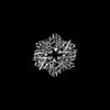
| Title | Structure of the MlaCD complex (1:6 stoichiometry) | |||||||||||||||||||||||||||||||||
 Components Components |
| |||||||||||||||||||||||||||||||||
 Keywords Keywords | LIPID BINDING PROTEIN / Outer membrane / gram-negative bacteria / lipid transfer / antibiotic resistance | |||||||||||||||||||||||||||||||||
| Function / homology |  Function and homology information Function and homology informationphospholipid transfer activity / intermembrane phospholipid transfer / phospholipid transport / phospholipid binding / outer membrane-bounded periplasmic space / membrane / plasma membrane Similarity search - Function | |||||||||||||||||||||||||||||||||
| Biological species |  | |||||||||||||||||||||||||||||||||
| Method | ELECTRON MICROSCOPY / single particle reconstruction / cryo EM / Resolution: 4.35 Å | |||||||||||||||||||||||||||||||||
 Authors Authors | Wotherspoon, P. / Bui, S. / Sridhar, P. / Bergeron, J.R.C. / Knowles, T.J. | |||||||||||||||||||||||||||||||||
| Funding support |  United Kingdom, 1items United Kingdom, 1items
| |||||||||||||||||||||||||||||||||
 Citation Citation | Journal: Nat Commun / Year: 2024 Title: Structure of the MlaC-MlaD complex reveals molecular basis of periplasmic phospholipid transport. Authors: Peter Wotherspoon / Hannah Johnston / David J Hardy / Rachel Holyfield / Soi Bui / Giedrė Ratkevičiūtė / Pooja Sridhar / Jonathan Colburn / Charlotte B Wilson / Adam Colyer / Benjamin F ...Authors: Peter Wotherspoon / Hannah Johnston / David J Hardy / Rachel Holyfield / Soi Bui / Giedrė Ratkevičiūtė / Pooja Sridhar / Jonathan Colburn / Charlotte B Wilson / Adam Colyer / Benjamin F Cooper / Jack A Bryant / Gareth W Hughes / Phillip J Stansfeld / Julien R C Bergeron / Timothy J Knowles / Abstract: The Maintenance of Lipid Asymmetry (Mla) pathway is a multicomponent system found in all gram-negative bacteria that contributes to virulence, vesicle blebbing and preservation of the outer membrane ...The Maintenance of Lipid Asymmetry (Mla) pathway is a multicomponent system found in all gram-negative bacteria that contributes to virulence, vesicle blebbing and preservation of the outer membrane barrier function. It acts by removing ectopic lipids from the outer leaflet of the outer membrane and returning them to the inner membrane through three proteinaceous assemblies: the MlaA-OmpC complex, situated within the outer membrane; the periplasmic phospholipid shuttle protein, MlaC; and the inner membrane ABC transporter complex, MlaFEDB, proposed to be the founding member of a structurally distinct ABC superfamily. While the function of each component is well established, how phospholipids are exchanged between components remains unknown. This stands as a major roadblock in our understanding of the function of the pathway, and in particular, the role of ATPase activity of MlaFEDB is not clear. Here, we report the structure of E. coli MlaC in complex with the MlaD hexamer in two distinct stoichiometries. Utilising in vivo complementation assays, an in vitro fluorescence-based transport assay, and molecular dynamics simulations, we confirm key residues, identifying the MlaD β6-β7 loop as essential for MlaCD function. We also provide evidence that phospholipids pass between the C-terminal helices of the MlaD hexamer to reach the central pore, providing insight into the trajectory of GPL transfer between MlaC and MlaD. | |||||||||||||||||||||||||||||||||
| History |
|
- Structure visualization
| Structure viewer | Molecule: MolmilJmol/JSmol |
|---|
- Downloads & links
-Download
| PDBx/mmCIF format | 8oj4.cif.gz | 167.6 KB | Display | PDBx/mmCIF format |
|---|---|---|---|---|
| PDB format | pdb8oj4.ent.gz | 130.2 KB | Display | PDB format |
| PDBx/mmJSON format | 8oj4.json.gz | Tree view | PDBx/mmJSON format | |
| Others |  Other downloads Other downloads |
-Validation report
| Arichive directory | https://data.pdbj.org/pub/pdb/validation_reports/oj/8oj4ftp://data.pdbj.org/pub/pdb/validation_reports/oj/8oj4 | HTTPS FTP |
|---|
-Related structure data
| Related structure data |  16904MC 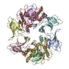 8ojgC M: map data used to model this data C: citing same article ( |
|---|---|
| Similar structure data |
-Links
 PDBj
PDBj- Assembly
| Deposited unit | 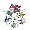
|
|---|---|
| 1 |
|
-Components
| #1: Protein | Mass: 23989.559 Da / Num. of mol.: 1 Source method: isolated from a genetically manipulated source Source: (gene. exp.) | ||
|---|---|---|---|
| #2: Protein | Mass: 19593.133 Da / Num. of mol.: 6 Source method: isolated from a genetically manipulated source Source: (gene. exp.) Has protein modification | N | |
-Experimental details
-Experiment
| Experiment | Method: ELECTRON MICROSCOPY |
|---|---|
| EM experiment | Aggregation state: PARTICLE / 3D reconstruction method: single particle reconstruction |
- Sample preparation
| Component | Name: MlaCD / Type: COMPLEX / Entity ID: all / Source: RECOMBINANT |
|---|---|
| Source (natural) | Organism: |
| Source (recombinant) | Organism: |
| Buffer solution | pH: 8 |
| Specimen | Embedding applied: NO / Shadowing applied: NO / Staining applied: NO / Vitrification applied: YES |
| Vitrification | Cryogen name: ETHANE |
- Electron microscopy imaging
| Experimental equipment |  Model: Titan Krios / Image courtesy: FEI Company |
|---|---|
| Microscopy | Model: FEI TITAN KRIOS |
| Electron gun | Electron source:  FIELD EMISSION GUN / Accelerating voltage: 300 kV / Illumination mode: FLOOD BEAM FIELD EMISSION GUN / Accelerating voltage: 300 kV / Illumination mode: FLOOD BEAM |
| Electron lens | Mode: BRIGHT FIELD / Nominal defocus max: 2000 nm / Nominal defocus min: 500 nm |
| Image recording | Electron dose: 40 e/Å2 / Film or detector model: GATAN K3 (6k x 4k) |
- Processing
| Software | Name: PHENIX / Version: 1.18rc3_3805: / Classification: refinement | ||||||||||||||||||||||||
|---|---|---|---|---|---|---|---|---|---|---|---|---|---|---|---|---|---|---|---|---|---|---|---|---|---|
| EM software | Name: PHENIX / Category: model refinement | ||||||||||||||||||||||||
| CTF correction | Type: PHASE FLIPPING AND AMPLITUDE CORRECTION | ||||||||||||||||||||||||
| 3D reconstruction | Resolution: 4.35 Å / Resolution method: FSC 0.143 CUT-OFF / Num. of particles: 97460 / Symmetry type: POINT | ||||||||||||||||||||||||
| Refine LS restraints |
|
