- EMDB-16904: Structure of the MlaCD complex (1:6 stoichiometry) -
+
Open data
ID or keywords:
Loading...
-
Basic information
Entry
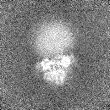
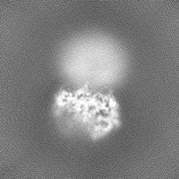
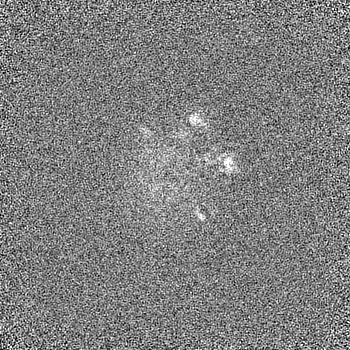
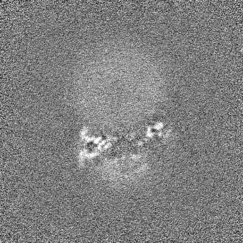
Database: EMDB / ID: EMD-16904
Title
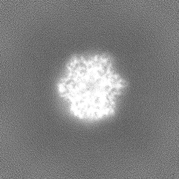
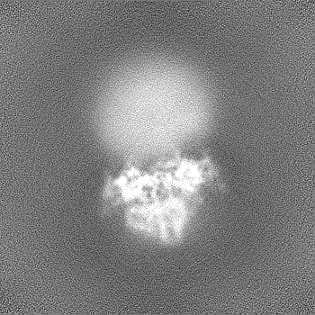
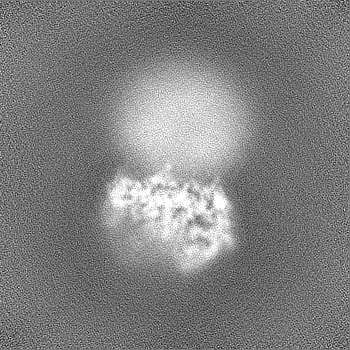
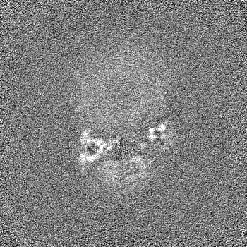
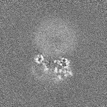
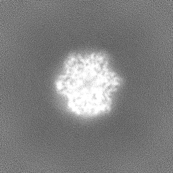
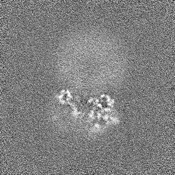
Structure of the MlaCD complex (1:6 stoichiometry)
Map data
Sample
Complex: MlaCD
Protein or peptide: Intermembrane phospholipid transport system binding protein MlaC
Protein or peptide: Intermembrane phospholipid transport system binding protein MlaD
Keywords
Outer membrane / gram-negative bacteria / lipid transfer / antibiotic resistance / LIPID BINDING PROTEIN
Function / homology
Function and homology information
phospholipid transfer activity / intermembrane phospholipid transfer / phospholipid transport / phospholipid binding / outer membrane-bounded periplasmic space / membrane / plasma membrane Similarity search - Function
Tgt2/MlaC superfamily / Toluene tolerance Ttg2/phospholipid-binding protein MlaC / MlaC protein / Probable phospholipid ABC transporter-binding protein MlaD / : / Mce/MlaD / MlaD protein Similarity search - Domain/homology
Intermembrane phospholipid transport system binding protein MlaC / Intermembrane phospholipid transport system binding protein MlaD Similarity search - Component
Biological species
Escherichia coli (E. coli)
Method
single particle reconstruction / cryo EM / Resolution: 4.35 Å
Biotechnology and Biological Sciences Research Council (BBSRC)
BB/R019061/2
United Kingdom
Citation
Journal: Nat Commun / Year: 2024 Title: Structure of the MlaC-MlaD complex reveals molecular basis of periplasmic phospholipid transport. Authors: Peter Wotherspoon / Hannah Johnston / David J Hardy / Rachel Holyfield / Soi Bui / Giedrė Ratkevičiūtė / Pooja Sridhar / Jonathan Colburn / Charlotte B Wilson / Adam Colyer / Benjamin F ...Authors: Peter Wotherspoon / Hannah Johnston / David J Hardy / Rachel Holyfield / Soi Bui / Giedrė Ratkevičiūtė / Pooja Sridhar / Jonathan Colburn / Charlotte B Wilson / Adam Colyer / Benjamin F Cooper / Jack A Bryant / Gareth W Hughes / Phillip J Stansfeld / Julien R C Bergeron / Timothy J Knowles / Abstract: The Maintenance of Lipid Asymmetry (Mla) pathway is a multicomponent system found in all gram-negative bacteria that contributes to virulence, vesicle blebbing and preservation of the outer membrane ...The Maintenance of Lipid Asymmetry (Mla) pathway is a multicomponent system found in all gram-negative bacteria that contributes to virulence, vesicle blebbing and preservation of the outer membrane barrier function. It acts by removing ectopic lipids from the outer leaflet of the outer membrane and returning them to the inner membrane through three proteinaceous assemblies: the MlaA-OmpC complex, situated within the outer membrane; the periplasmic phospholipid shuttle protein, MlaC; and the inner membrane ABC transporter complex, MlaFEDB, proposed to be the founding member of a structurally distinct ABC superfamily. While the function of each component is well established, how phospholipids are exchanged between components remains unknown. This stands as a major roadblock in our understanding of the function of the pathway, and in particular, the role of ATPase activity of MlaFEDB is not clear. Here, we report the structure of E. coli MlaC in complex with the MlaD hexamer in two distinct stoichiometries. Utilising in vivo complementation assays, an in vitro fluorescence-based transport assay, and molecular dynamics simulations, we confirm key residues, identifying the MlaD β6-β7 loop as essential for MlaCD function. We also provide evidence that phospholipids pass between the C-terminal helices of the MlaD hexamer to reach the central pore, providing insight into the trajectory of GPL transfer between MlaC and MlaD.
In the structure databanks used in Yorodumi, some data are registered as the other names, "COVID-19 virus" and "2019-nCoV". Here are the details of the virus and the list of structure data.
Jan 31, 2019. EMDB accession codes are about to change! (news from PDBe EMDB page)
EMDB accession codes are about to change! (news from PDBe EMDB page)
The allocation of 4 digits for EMDB accession codes will soon come to an end. Whilst these codes will remain in use, new EMDB accession codes will include an additional digit and will expand incrementally as the available range of codes is exhausted. The current 4-digit format prefixed with “EMD-” (i.e. EMD-XXXX) will advance to a 5-digit format (i.e. EMD-XXXXX), and so on. It is currently estimated that the 4-digit codes will be depleted around Spring 2019, at which point the 5-digit format will come into force.
The EM Navigator/Yorodumi systems omit the EMD- prefix.
Related info.:Q: What is EMD? / ID/Accession-code notation in Yorodumi/EM Navigator
Yorodumi is a browser for structure data from EMDB, PDB, SASBDB, etc.
This page is also the successor to EM Navigator detail page, and also detail information page/front-end page for Omokage search.
The word "yorodu" (or yorozu) is an old Japanese word meaning "ten thousand". "mi" (miru) is to see.
Related info.:EMDB / PDB / SASBDB / Comparison of 3 databanks / Yorodumi Search / Aug 31, 2016. New EM Navigator & Yorodumi / Yorodumi Papers / Jmol/JSmol / Function and homology information / Changes in new EM Navigator and Yorodumi
 Movie
Movie Controller


 Open data
 Basic information
 Map data
 Sample
 Keywords
 Function and homology information
 Authors
 United Kingdom, 1 items
  Citation
 Structure visualization
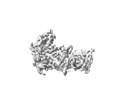
 Downloads & links
 emd_16904.png
 http://ftp.pdbj.org/pub/emdb/structures/EMD-16904
 Z (Sec.)
 Y (Row.)
 X (Col.)


 Sample components
 Processing
 Electron microscopy
 FIELD EMISSION GUN
