 Movie
Movie Controller

[English] 日本語
 Yorodumi
- PDB-8ofw: Crystal structure of the full-length dihydroorotate dehydrogenase... -
+ Open data

- Basic information
| Entry | Database: PDB / ID: 8ofw | ||||||
|---|---|---|---|---|---|---|---|
| Title | Crystal structure of the full-length dihydroorotate dehydrogenase from Mycobacterium tuberculosis | ||||||
 Components Components | Dihydroorotate dehydrogenase (quinone) | ||||||
 Keywords Keywords | OXIDOREDUCTASE / dihydroorotate dehydrogenase / Flavoenzyme / Pyrimidine biosynthesis | ||||||
| Function / homology |  Function and homology information Function and homology informationpyrimidine ribonucleotide biosynthetic process / dihydroorotate dehydrogenase (quinone) / dihydroorotate dehydrogenase (quinone) activity / dihydroorotate dehydrogenase activity / 'de novo' UMP biosynthetic process / 'de novo' pyrimidine nucleobase biosynthetic process / plasma membrane / cytoplasm Similarity search - Function | ||||||
| Biological species |   Mycobacterium tuberculosis (bacteria) Mycobacterium tuberculosis (bacteria) | ||||||
| Method |  X-RAY DIFFRACTION / SYNCHROTRON / MOLECULAR REPLACEMENT / Resolution: 3.8 Å X-RAY DIFFRACTION / SYNCHROTRON / MOLECULAR REPLACEMENT / Resolution: 3.8 Å | ||||||
 Authors Authors | Alberti, M. / Ferraris, D.M. / Miggiano, R. | ||||||
| Funding support |  Italy, 1items Italy, 1items
| ||||||
 Citation Citation | Journal: Febs Lett. / Year: 2023 Title: Biochemical characterization of Mycobacterium tuberculosis dihydroorotate dehydrogenase and identification of a selective inhibitor. Authors: Alberti, M. / Sainas, S. / Ronchi, E. / Lolli, M.L. / Boschi, D. / Rizzi, M. / Ferraris, D.M. / Miggiano, R. | ||||||
| History |
|
- Structure visualization
| Structure viewer | Molecule: MolmilJmol/JSmol |
|---|
- Downloads & links
-Download
| PDBx/mmCIF format | 8ofw.cif.gz | 149.5 KB | Display | PDBx/mmCIF format |
|---|---|---|---|---|
| PDB format | pdb8ofw.ent.gz | 111.3 KB | Display | PDB format |
| PDBx/mmJSON format | 8ofw.json.gz | Tree view | PDBx/mmJSON format | |
| Others |  Other downloads Other downloads |
-Validation report
| Arichive directory | https://data.pdbj.org/pub/pdb/validation_reports/of/8ofwftp://data.pdbj.org/pub/pdb/validation_reports/of/8ofw | HTTPS FTP |
|---|
-Related structure data
| Similar structure data |
|---|
-Links
 PDBj
PDBj- Assembly
| Deposited unit | 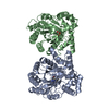
| ||||||||||
|---|---|---|---|---|---|---|---|---|---|---|---|
| 1 | 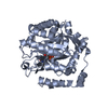
| ||||||||||
| 2 | 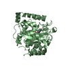
| ||||||||||
| Unit cell |
| ||||||||||
| Noncrystallographic symmetry (NCS) | NCS oper: (Code: givenMatrix: (-0.999321541726, -0.0309324818799, 0.0199909431206), (0.0284020368173, -0.992796540085, -0.116397398167), (0.0234473995728, -0.115750643887, 0.993001514547)Vector: -23. ...NCS oper: (Code: given Matrix: (-0.999321541726, -0.0309324818799, 0.0199909431206), Vector: |
-Components
| #1: Protein | Mass: 40219.934 Da / Num. of mol.: 2 Source method: isolated from a genetically manipulated source Source: (gene. exp.) Mycobacterium tuberculosis (bacteria) / Gene: pyrD, Rv2139, MTCY270.29c / Production host: References: UniProt: P9WHL1, dihydroorotate dehydrogenase (quinone) #2: Chemical |   Mass: 456.344 Da / Num. of mol.: 2 / Source method: obtained synthetically / Formula: C17H21N4O9P / Feature type: SUBJECT OF INVESTIGATION Mass: 456.344 Da / Num. of mol.: 2 / Source method: obtained synthetically / Formula: C17H21N4O9P / Feature type: SUBJECT OF INVESTIGATIONHas ligand of interest | Y | |
|---|
-Experimental details
-Experiment
| Experiment | Method: X-RAY DIFFRACTION / Number of used crystals: 1 |
|---|
- Sample preparation
| Crystal | Density Matthews: 2.21 Å3/Da / Density % sol: 44.24 % |
|---|---|
| Crystal grow | Temperature: 293.15 K / Method: vapor diffusion, hanging drop / pH: 8.5 Details: 0,2 M Sodium Chloride, 0,1 M Tris-HCl pH 8,5 and 25 % (w/v) PEG 3350 |
-Data collection
| Diffraction | Mean temperature: 100 K / Serial crystal experiment: N |
|---|---|
| Diffraction source | Source: SYNCHROTRON / Site: ESRF  / Beamline: ID23-2 / Wavelength: 0.873128 Å / Beamline: ID23-2 / Wavelength: 0.873128 Å |
| Detector | Type: DECTRIS PILATUS3 2M / Detector: PIXEL / Date: Jan 27, 2022 |
| Radiation | Protocol: SINGLE WAVELENGTH / Monochromatic (M) / Laue (L): M / Scattering type: x-ray |
| Radiation wavelength | Wavelength: 0.873128 Å / Relative weight: 1 |
| Reflection | Resolution: 3.8→41.508 Å / Num. obs: 39498 / % possible obs: 97.5 % / Redundancy: 5.5 % / Biso Wilson estimate: 116.1 Å2 / CC1/2: 0.988 / Rmerge(I) obs: 0.253 / Rrim(I) all: 0.279 / Net I/σ(I): 5 |
| Reflection shell | Resolution: 3.8→4 Å / Rmerge(I) obs: 1.229 / Mean I/σ(I) obs: 1.5 / Num. unique obs: 1012 / CC1/2: 0.688 / Rrim(I) all: 1.358 |
- Processing
| Software |
| |||||||||||||||||||||||||||||||||||||||||||||||||||||||||||||||||||||||||||||||||||||||||||||||||||||||||||||||||||||||||||||||||||||||||||||||||||||||||||||||||||||||||||||||||||||||||||||||||||||||||||||||||||||||||||||||||||||||
|---|---|---|---|---|---|---|---|---|---|---|---|---|---|---|---|---|---|---|---|---|---|---|---|---|---|---|---|---|---|---|---|---|---|---|---|---|---|---|---|---|---|---|---|---|---|---|---|---|---|---|---|---|---|---|---|---|---|---|---|---|---|---|---|---|---|---|---|---|---|---|---|---|---|---|---|---|---|---|---|---|---|---|---|---|---|---|---|---|---|---|---|---|---|---|---|---|---|---|---|---|---|---|---|---|---|---|---|---|---|---|---|---|---|---|---|---|---|---|---|---|---|---|---|---|---|---|---|---|---|---|---|---|---|---|---|---|---|---|---|---|---|---|---|---|---|---|---|---|---|---|---|---|---|---|---|---|---|---|---|---|---|---|---|---|---|---|---|---|---|---|---|---|---|---|---|---|---|---|---|---|---|---|---|---|---|---|---|---|---|---|---|---|---|---|---|---|---|---|---|---|---|---|---|---|---|---|---|---|---|---|---|---|---|---|---|---|---|---|---|---|---|---|---|---|---|---|---|---|---|---|---|---|
| Refinement | Method to determine structure: MOLECULAR REPLACEMENT / Resolution: 3.8→41.508 Å / Cor.coef. Fo:Fc: 0.913 / Cor.coef. Fo:Fc free: 0.856 / WRfactor Rfree: 0.307 / WRfactor Rwork: 0.235 / SU B: 92.873 / SU ML: 1.209 / Average fsc free: 0.9077 / Average fsc work: 0.9303 / Cross valid method: FREE R-VALUE / ESU R Free: 1.126 Details: Hydrogens have been added in their riding positions
| |||||||||||||||||||||||||||||||||||||||||||||||||||||||||||||||||||||||||||||||||||||||||||||||||||||||||||||||||||||||||||||||||||||||||||||||||||||||||||||||||||||||||||||||||||||||||||||||||||||||||||||||||||||||||||||||||||||||
| Solvent computation | Ion probe radii: 0.8 Å / Shrinkage radii: 0.8 Å / VDW probe radii: 1.2 Å / Solvent model: MASK BULK SOLVENT | |||||||||||||||||||||||||||||||||||||||||||||||||||||||||||||||||||||||||||||||||||||||||||||||||||||||||||||||||||||||||||||||||||||||||||||||||||||||||||||||||||||||||||||||||||||||||||||||||||||||||||||||||||||||||||||||||||||||
| Displacement parameters | Biso mean: 125.462 Å2
| |||||||||||||||||||||||||||||||||||||||||||||||||||||||||||||||||||||||||||||||||||||||||||||||||||||||||||||||||||||||||||||||||||||||||||||||||||||||||||||||||||||||||||||||||||||||||||||||||||||||||||||||||||||||||||||||||||||||
| Refinement step | Cycle: LAST / Resolution: 3.8→41.508 Å
| |||||||||||||||||||||||||||||||||||||||||||||||||||||||||||||||||||||||||||||||||||||||||||||||||||||||||||||||||||||||||||||||||||||||||||||||||||||||||||||||||||||||||||||||||||||||||||||||||||||||||||||||||||||||||||||||||||||||
| Refine LS restraints |
| |||||||||||||||||||||||||||||||||||||||||||||||||||||||||||||||||||||||||||||||||||||||||||||||||||||||||||||||||||||||||||||||||||||||||||||||||||||||||||||||||||||||||||||||||||||||||||||||||||||||||||||||||||||||||||||||||||||||
| LS refinement shell | Refine-ID: X-RAY DIFFRACTION / Total num. of bins used: 20
|
