 Movie
Movie Controller

[English] 日本語
 Yorodumi
- PDB-8ofs: Human adenovirus type 30 fiber-knob protein complexed with sialic acid -
+ Open data

- Basic information
| Entry | Database: PDB / ID: 8ofs | |||||||||
|---|---|---|---|---|---|---|---|---|---|---|
| Title | Human adenovirus type 30 fiber-knob protein complexed with sialic acid | |||||||||
 Components Components | Fiber | |||||||||
 Keywords Keywords | VIRAL PROTEIN / Adenovirus / Fiber knob / Ad25 | |||||||||
| Function / homology |  Function and homology information Function and homology informationadhesion receptor-mediated virion attachment to host cell / viral capsid / cell adhesion / symbiont entry into host cell / host cell nucleus Similarity search - Function | |||||||||
| Biological species |  Human adenovirus 30 Human adenovirus 30 | |||||||||
| Method |  X-RAY DIFFRACTION / SYNCHROTRON / MOLECULAR REPLACEMENT / molecular replacement / Resolution: 2.56 Å X-RAY DIFFRACTION / SYNCHROTRON / MOLECULAR REPLACEMENT / molecular replacement / Resolution: 2.56 Å | |||||||||
 Authors Authors | Rizkallah, P.J. / Parker, A.L. / Mundy, R.M. / Baker, A.T. | |||||||||
| Funding support |  United Kingdom, 2items United Kingdom, 2items
| |||||||||
 Citation Citation | Journal: Npj Viruses / Year: 2023 Title: Broad sialic acid usage amongst species D human adenovirus. Authors: Mundy, R.M. / Baker, A.T. / Bates, E.A. / Cunliffe, T.G. / Teijeira-Crespo, A. / Moses, E. / Rizkallah, P.J. / Parker, A.L. | |||||||||
| History |
|
- Structure visualization
| Structure viewer | Molecule: MolmilJmol/JSmol |
|---|
- Downloads & links
-Download
| PDBx/mmCIF format | 8ofs.cif.gz | 254.8 KB | Display | PDBx/mmCIF format |
|---|---|---|---|---|
| PDB format | pdb8ofs.ent.gz | 209.2 KB | Display | PDB format |
| PDBx/mmJSON format | 8ofs.json.gz | Tree view | PDBx/mmJSON format | |
| Others |  Other downloads Other downloads |
-Validation report
| Summary document | 8ofs_validation.pdf.gz | 1.4 MB | Display | wwPDB validaton report |
|---|---|---|---|---|
| Full document | 8ofs_full_validation.pdf.gz | 1.4 MB | Display | |
| Data in XML | 8ofs_validation.xml.gz | 24.5 KB | Display | |
| Data in CIF | 8ofs_validation.cif.gz | 31.5 KB | Display | |
| Arichive directory | https://data.pdbj.org/pub/pdb/validation_reports/of/8ofsftp://data.pdbj.org/pub/pdb/validation_reports/of/8ofs | HTTPS FTP |
-Related structure data
| Related structure data |  8ofpC  8ofqC  8ofrC  8oftC 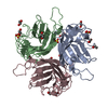 8ofuC  8ofvC C: citing same article ( |
|---|---|
| Similar structure data |
-Links
 PDBj
PDBj
- Assembly
| Deposited unit | 
| ||||||||||||
|---|---|---|---|---|---|---|---|---|---|---|---|---|---|
| 1 |
| ||||||||||||
| Unit cell |
| ||||||||||||
| Noncrystallographic symmetry (NCS) | NCS domain:
|
-Components
| #1: Protein | Mass: 22290.828 Da / Num. of mol.: 3 Source method: isolated from a genetically manipulated source Source: (gene. exp.) Human adenovirus 30 / Strain: 30 / Gene: L5 / Production host:  #2: Sugar |   Type: D-saccharide, beta linking / Mass: 309.270 Da / Num. of mol.: 3 / Source method: obtained synthetically / Formula: C11H19NO9 / Feature type: SUBJECT OF INVESTIGATION Type: D-saccharide, beta linking / Mass: 309.270 Da / Num. of mol.: 3 / Source method: obtained synthetically / Formula: C11H19NO9 / Feature type: SUBJECT OF INVESTIGATIONHas ligand of interest | Y | Has protein modification | Y | |
|---|
-Experimental details
-Experiment
| Experiment | Method: X-RAY DIFFRACTION / Number of used crystals: 1 |
|---|
- Sample preparation
| Crystal | Density Matthews: 2.07 Å3/Da / Density % sol: 40.67 % |
|---|---|
| Crystal grow | Temperature: 291 K / Method: vapor diffusion, sitting drop / pH: 8 / Details: 0.1M MMT [Malic acid, MES, Tris], 25% w/v PEG 1500 |
-Data collection
| Diffraction | Mean temperature: 100 K / Serial crystal experiment: N |
|---|---|
| Diffraction source | Source: SYNCHROTRON / Site: Diamond / Beamline: I04 / Wavelength: 0.9795 Å |
| Detector | Type: DECTRIS EIGER X 16M / Detector: PIXEL / Date: May 18, 2019 |
| Radiation | Protocol: SINGLE WAVELENGTH / Monochromatic (M) / Laue (L): M / Scattering type: x-ray |
| Radiation wavelength | Wavelength: 0.9795 Å / Relative weight: 1 |
| Reflection | Resolution: 2.56→62.49 Å / Num. obs: 17735 / % possible obs: 99 % / Redundancy: 3.7 % / Biso Wilson estimate: 74.3 Å2 / CC1/2: 0.998 / Rmerge(I) obs: 0.057 / Rpim(I) all: 0.034 / Rrim(I) all: 0.066 / Χ2: 0.95 / Net I/σ(I): 10.5 |
| Reflection shell | Resolution: 2.56→2.63 Å / % possible obs: 99.3 % / Redundancy: 3.8 % / Rmerge(I) obs: 0.972 / Num. measured all: 5020 / Num. unique obs: 1327 / CC1/2: 0.824 / Rpim(I) all: 0.57 / Rrim(I) all: 1.131 / Χ2: 0.87 / Net I/σ(I) obs: 1.1 |
-Phasing
| Phasing | Method: molecular replacement | ||||||
|---|---|---|---|---|---|---|---|
| Phasing MR | Model details: Phaser MODE: MR_AUTO
|
- Processing
| Software |
| ||||||||||||||||||||||||||||||||||||||||||||||||||||||||||||||||||||||||||||||||||||||||||||||||||||||||||||||||||||||||||||||||||||||||||||||||||||||||||||||||||||||||||||||||||||||
|---|---|---|---|---|---|---|---|---|---|---|---|---|---|---|---|---|---|---|---|---|---|---|---|---|---|---|---|---|---|---|---|---|---|---|---|---|---|---|---|---|---|---|---|---|---|---|---|---|---|---|---|---|---|---|---|---|---|---|---|---|---|---|---|---|---|---|---|---|---|---|---|---|---|---|---|---|---|---|---|---|---|---|---|---|---|---|---|---|---|---|---|---|---|---|---|---|---|---|---|---|---|---|---|---|---|---|---|---|---|---|---|---|---|---|---|---|---|---|---|---|---|---|---|---|---|---|---|---|---|---|---|---|---|---|---|---|---|---|---|---|---|---|---|---|---|---|---|---|---|---|---|---|---|---|---|---|---|---|---|---|---|---|---|---|---|---|---|---|---|---|---|---|---|---|---|---|---|---|---|---|---|---|---|
| Refinement | Method to determine structure: MOLECULAR REPLACEMENT / Resolution: 2.56→48.56 Å / Cor.coef. Fo:Fc: 0.968 / Cor.coef. Fo:Fc free: 0.936 / SU B: 107.9 / SU ML: 0.768 / Cross valid method: THROUGHOUT / ESU R Free: 0.405 / Stereochemistry target values: MAXIMUM LIKELIHOOD / Details: HYDROGENS HAVE BEEN ADDED IN THE RIDING POSITIONS
| ||||||||||||||||||||||||||||||||||||||||||||||||||||||||||||||||||||||||||||||||||||||||||||||||||||||||||||||||||||||||||||||||||||||||||||||||||||||||||||||||||||||||||||||||||||||
| Solvent computation | Ion probe radii: 0.8 Å / Shrinkage radii: 0.8 Å / VDW probe radii: 1.2 Å / Solvent model: MASK | ||||||||||||||||||||||||||||||||||||||||||||||||||||||||||||||||||||||||||||||||||||||||||||||||||||||||||||||||||||||||||||||||||||||||||||||||||||||||||||||||||||||||||||||||||||||
| Displacement parameters | Biso mean: 130.013 Å2
| ||||||||||||||||||||||||||||||||||||||||||||||||||||||||||||||||||||||||||||||||||||||||||||||||||||||||||||||||||||||||||||||||||||||||||||||||||||||||||||||||||||||||||||||||||||||
| Refinement step | Cycle: 1 / Resolution: 2.56→48.56 Å
| ||||||||||||||||||||||||||||||||||||||||||||||||||||||||||||||||||||||||||||||||||||||||||||||||||||||||||||||||||||||||||||||||||||||||||||||||||||||||||||||||||||||||||||||||||||||
| Refine LS restraints |
|