Movie
Movie Controller

[English] 日本語
 Yorodumi
- PDB-8of8: Cryo-EM structure of actomyosin-5a-S1 with the full-length lever ... -
+ Open data

- Basic information
| Entry | Database: PDB / ID: 8of8 | ||||||||||||||||||
|---|---|---|---|---|---|---|---|---|---|---|---|---|---|---|---|---|---|---|---|
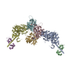
| Title | Cryo-EM structure of actomyosin-5a-S1 with the full-length lever (nucleotide free) | ||||||||||||||||||
 Components Components |
| ||||||||||||||||||
 Keywords Keywords | MOTOR PROTEIN / myosin / cytoskeletal motor / myosin-va / myo5a / myosin-5a / S1 / rigor / nucleotide free / apo / lever / 6IQ / actomyosin / actomyosin-5a / actin / actomyosin-va / actin bound | ||||||||||||||||||
| Function / homology |  Function and homology information Function and homology informationactomyosin, myosin complex part / establishment of endoplasmic reticulum localization to postsynapse / axo-dendritic protein transport / regulation of postsynaptic cytosolic calcium ion concentration / positive regulation of cellular response to insulin stimulus / CaMK IV-mediated phosphorylation of CREB / Cam-PDE 1 activation / CREB1 phosphorylation through the activation of CaMKII/CaMKK/CaMKIV cascasde / Glycogen breakdown (glycogenolysis) / Activation of RAC1 downstream of NMDARs ...actomyosin, myosin complex part / establishment of endoplasmic reticulum localization to postsynapse / axo-dendritic protein transport / regulation of postsynaptic cytosolic calcium ion concentration / positive regulation of cellular response to insulin stimulus / CaMK IV-mediated phosphorylation of CREB / Cam-PDE 1 activation / CREB1 phosphorylation through the activation of CaMKII/CaMKK/CaMKIV cascasde / Glycogen breakdown (glycogenolysis) / Activation of RAC1 downstream of NMDARs / Sodium/Calcium exchangers / Activation of Ca-permeable Kainate Receptor / melanosome localization / CLEC7A (Dectin-1) induces NFAT activation / RHO GTPases activate PAKs / Calmodulin induced events / Synthesis of IP3 and IP4 in the cytosol / Inactivation, recovery and regulation of the phototransduction cascade / Tetrahydrobiopterin (BH4) synthesis, recycling, salvage and regulation / eNOS activation / endoplasmic reticulum localization / Reduction of cytosolic Ca++ levels / locomotion involved in locomotory behavior / Calcineurin activates NFAT / Ion transport by P-type ATPases / RAF activation / VEGFR2 mediated vascular permeability / melanin metabolic process / Protein methylation / vesicle transport along actin filament / negative regulation of dopamine secretion / RAS processing / insulin-responsive compartment / Ca2+ pathway / Extra-nuclear estrogen signaling / FCERI mediated Ca+2 mobilization / RHO GTPases activate IQGAPs / Unblocking of NMDA receptors, glutamate binding and activation / post-Golgi vesicle-mediated transport / unconventional myosin complex / PKA activation / secretory granule localization / RAF/MAP kinase cascade / developmental pigmentation / reactive gliosis / Regulation of MITF-M-dependent genes involved in pigmentation / Regulation of actin dynamics for phagocytic cup formation / Smooth Muscle Contraction / filopodium tip / Platelet degranulation / melanin biosynthetic process / High laminar flow shear stress activates signaling by PIEZO1 and PECAM1:CDH5:KDR in endothelial cells / regulation of exocytosis / melanocyte differentiation / hair follicle maturation / actin filament-based movement / actomyosin / Stimuli-sensing channels / melanosome transport / : / Ion homeostasis / type 3 metabotropic glutamate receptor binding / postsynaptic actin cytoskeleton / ATP-dependent protein binding / positive regulation of vascular associated smooth muscle cell migration / myosin complex / odontogenesis / long-chain fatty acid biosynthetic process / syntaxin-1 binding / negative regulation of synaptic transmission, glutamatergic / insulin secretion / intermediate filament / cytoskeletal motor activator activity / microfilament motor activity / response to corticosterone / negative regulation of ryanodine-sensitive calcium-release channel activity / organelle localization by membrane tethering / myosin heavy chain binding / : / autophagosome membrane docking / regulation of synaptic vesicle exocytosis / regulation of ryanodine-sensitive calcium-release channel activity / regulation of cardiac muscle cell action potential / tropomyosin binding / presynaptic endocytosis / actin filament bundle / troponin I binding / smooth endoplasmic reticulum / dopamine metabolic process / pigmentation / filamentous actin / exocytosis / mesenchyme migration / calcineurin-mediated signaling / nitric-oxide synthase binding / cytoskeletal motor activity / regulation of cell communication by electrical coupling involved in cardiac conduction / skeletal muscle myofibril / actin filament bundle assembly / adenylate cyclase binding Similarity search - Function | ||||||||||||||||||
| Biological species |  | ||||||||||||||||||
| Method | ELECTRON MICROSCOPY / single particle reconstruction / cryo EM / Resolution: 7.5 Å | ||||||||||||||||||
 Authors Authors | Gravett, M.S.C. / Klebl, D.P. / Harlen, O.G. / Read, D.J. / Harris, S.A. / Muench, S.P. / Peckham, M. | ||||||||||||||||||
| Funding support |  United Kingdom, 5items United Kingdom, 5items
| ||||||||||||||||||
 Citation Citation | Journal: Structure / Year: 2024 Title: Exploiting cryo-EM structures of actomyosin-5a to reveal the physical properties of its lever. Authors: Molly S C Gravett / David P Klebl / Oliver G Harlen / Daniel J Read / Stephen P Muench / Sarah A Harris / Michelle Peckham / Abstract: Myosin 5a (Myo5a) is a dimeric processive motor protein that transports cellular cargos along filamentous actin (F-actin). Its long lever is responsible for its large power-stroke, step size, and ...Myosin 5a (Myo5a) is a dimeric processive motor protein that transports cellular cargos along filamentous actin (F-actin). Its long lever is responsible for its large power-stroke, step size, and load-bearing ability. Little is known about the levers' structure and physical properties, and how they contribute to walking mechanics. Using cryoelectron microscopy (cryo-EM) and molecular dynamics (MD) simulations, we resolved the structure of monomeric Myo5a, comprising the motor domain and full-length lever, bound to F-actin. The range of its lever conformations revealed its physical properties, how stiffness varies along its length and predicts a large, 35 nm, working stroke. Thus, the newly released trail head in a dimeric Myo5a would only need to perform a small diffusive search for its new binding site on F-actin, and stress would only be generated across the dimer once phosphate is released from the lead head, revealing new insight into the walking behavior of Myo5a. | ||||||||||||||||||
| History |
|
- Structure visualization
| Structure viewer | Molecule: MolmilJmol/JSmol |
|---|
- Downloads & links
-Download
| PDBx/mmCIF format | 8of8.cif.gz | 410 KB | Display | PDBx/mmCIF format |
|---|---|---|---|---|
| PDB format | pdb8of8.ent.gz | 282.3 KB | Display | PDB format |
| PDBx/mmJSON format | 8of8.json.gz | Tree view | PDBx/mmJSON format | |
| Others |  Other downloads Other downloads |
-Validation report
| Arichive directory | https://data.pdbj.org/pub/pdb/validation_reports/of/8of8ftp://data.pdbj.org/pub/pdb/validation_reports/of/8of8 | HTTPS FTP |
|---|
-Related structure data
| Related structure data | 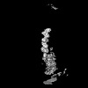 16850MC M: map data used to model this data C: citing same article ( |
|---|---|
| Similar structure data |


-Links
 PDBj
PDBj


- Assembly
| Deposited unit | 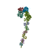
|
|---|---|
| 1 |
|
-Components
| #1: Protein | Mass: 16721.350 Da / Num. of mol.: 6 Source method: isolated from a genetically manipulated source Source: (gene. exp.)   Spodoptera frugiperda (fall armyworm) / References: UniProt: P0DP26 Spodoptera frugiperda (fall armyworm) / References: UniProt: P0DP26#2: Protein | Mass: 41875.633 Da / Num. of mol.: 3 / Source method: isolated from a natural source / Details: HIC is tele-methylhistidine / Source: (natural) #3: Protein | | Mass: 106596.195 Da / Num. of mol.: 1 Source method: isolated from a genetically manipulated source Source: (gene. exp.) Spodoptera frugiperda (fall armyworm) / References: UniProt: Q99104Has ligand of interest | N | Has protein modification | Y | |
|---|
-Experimental details
-Experiment
| Experiment | Method: ELECTRON MICROSCOPY |
|---|---|
| EM experiment | Aggregation state: HELICAL ARRAY / 3D reconstruction method: single particle reconstruction |
- Sample preparation
| Component |
| |||||||||||||||||||||||||||||||||||
|---|---|---|---|---|---|---|---|---|---|---|---|---|---|---|---|---|---|---|---|---|---|---|---|---|---|---|---|---|---|---|---|---|---|---|---|---|
| Molecular weight |
| |||||||||||||||||||||||||||||||||||
| Source (natural) |
| |||||||||||||||||||||||||||||||||||
| Source (recombinant) |
| |||||||||||||||||||||||||||||||||||
| Buffer solution | pH: 7 | |||||||||||||||||||||||||||||||||||
| Buffer component |
| |||||||||||||||||||||||||||||||||||
| Specimen | Embedding applied: NO / Shadowing applied: NO / Staining applied: NO / Vitrification applied: YES Details: 5.26 mg/mL Actin, alpha skeletal muscle 266 mg/mL FlAG-tagged unconventional myosin-Va (residues 1-907) 84.2 mg/mL Calmodulin-1 | |||||||||||||||||||||||||||||||||||
| Specimen support | Grid material: COPPER / Grid mesh size: 300 divisions/in. / Grid type: Quantifoil R2/2 | |||||||||||||||||||||||||||||||||||
| Vitrification | Instrument: FEI VITROBOT MARK IV / Cryogen name: ETHANE / Humidity: 80 % / Chamber temperature: 281.15 K |
- Electron microscopy imaging
| Experimental equipment |  Model: Titan Krios / Image courtesy: FEI Company |
|---|---|
| Microscopy | Model: TFS KRIOS |
| Electron gun | Electron source:  FIELD EMISSION GUN / Accelerating voltage: 300 kV / Illumination mode: FLOOD BEAM FIELD EMISSION GUN / Accelerating voltage: 300 kV / Illumination mode: FLOOD BEAM |
| Electron lens | Mode: BRIGHT FIELD / Nominal magnification: 75000 X / Nominal defocus max: 3600 nm / Nominal defocus min: 1800 nm / Cs: 2.7 mm / C2 aperture diameter: 50 µm / Alignment procedure: COMA FREE |
| Specimen holder | Cryogen: NITROGEN / Specimen holder model: FEI TITAN KRIOS AUTOGRID HOLDER |
| Image recording | Average exposure time: 1.5 sec. / Electron dose: 63.13 e/Å2 / Detector mode: INTEGRATING / Film or detector model: FEI FALCON III (4k x 4k) / Num. of grids imaged: 1 / Num. of real images: 4285 |
- Processing
| EM software |
| |||||||||||||||||||||||||||||||||||||||||||||
|---|---|---|---|---|---|---|---|---|---|---|---|---|---|---|---|---|---|---|---|---|---|---|---|---|---|---|---|---|---|---|---|---|---|---|---|---|---|---|---|---|---|---|---|---|---|---|
| CTF correction | Type: PHASE FLIPPING AND AMPLITUDE CORRECTION | |||||||||||||||||||||||||||||||||||||||||||||
| Particle selection | Num. of particles selected: 838213 | |||||||||||||||||||||||||||||||||||||||||||||
| Symmetry | Point symmetry: C1 (asymmetric) | |||||||||||||||||||||||||||||||||||||||||||||
| 3D reconstruction | Resolution: 7.5 Å / Resolution method: FSC 0.143 CUT-OFF / Num. of particles: 22337 / Algorithm: FOURIER SPACE / Num. of class averages: 1 / Symmetry type: POINT | |||||||||||||||||||||||||||||||||||||||||||||
| Atomic model building | Protocol: FLEXIBLE FIT / Space: REAL | |||||||||||||||||||||||||||||||||||||||||||||
| Atomic model building | 3D fitting-ID: 1
|