 Movie
Movie Controller

[English] 日本語
 Yorodumi
- EMDB-16846: Cryo-EM structure of actomyosin-5a-S1 with the full-length lever ... -
+ Open data

- Basic information
| Entry |  | ||||||||||||||||||
|---|---|---|---|---|---|---|---|---|---|---|---|---|---|---|---|---|---|---|---|
| Title | Cryo-EM structure of actomyosin-5a-S1 with the full-length lever (nucleotide free, class A) | ||||||||||||||||||
 Map data Map data | Main map, DeepEMhancer post-processed | ||||||||||||||||||
 Sample Sample |
| ||||||||||||||||||
 Keywords Keywords | myosin / cytoskeletal motor / myosin-va / myo5a / myosin-5a / S1 / rigor / nucleotide free / apo / lever / 6IQ / actomyosin / actomyosin-5a / actin / actomyosin-va / actin bound / MOTOR PROTEIN | ||||||||||||||||||
| Biological species |  | ||||||||||||||||||
| Method | single particle reconstruction / cryo EM / Resolution: 8.6 Å | ||||||||||||||||||
 Authors Authors | Gravett MSC / Klebl DP / Harlen OG / Read DJ / Harris SA / Muench SP / Peckham M | ||||||||||||||||||
| Funding support |  United Kingdom, 5 items United Kingdom, 5 items
| ||||||||||||||||||
 Citation Citation | Journal: Structure / Year: 2024 Title: Exploiting cryo-EM structures of actomyosin-5a to reveal the physical properties of its lever. Authors: Molly S C Gravett / David P Klebl / Oliver G Harlen / Daniel J Read / Stephen P Muench / Sarah A Harris / Michelle Peckham / Abstract: Myosin 5a (Myo5a) is a dimeric processive motor protein that transports cellular cargos along filamentous actin (F-actin). Its long lever is responsible for its large power-stroke, step size, and ...Myosin 5a (Myo5a) is a dimeric processive motor protein that transports cellular cargos along filamentous actin (F-actin). Its long lever is responsible for its large power-stroke, step size, and load-bearing ability. Little is known about the levers' structure and physical properties, and how they contribute to walking mechanics. Using cryoelectron microscopy (cryo-EM) and molecular dynamics (MD) simulations, we resolved the structure of monomeric Myo5a, comprising the motor domain and full-length lever, bound to F-actin. The range of its lever conformations revealed its physical properties, how stiffness varies along its length and predicts a large, 35 nm, working stroke. Thus, the newly released trail head in a dimeric Myo5a would only need to perform a small diffusive search for its new binding site on F-actin, and stress would only be generated across the dimer once phosphate is released from the lead head, revealing new insight into the walking behavior of Myo5a. | ||||||||||||||||||
| History |
|
- Structure visualization
| Supplemental images |
|---|

- Downloads & links
-EMDB archive
| Map data | emd_16846.map.gz | 55.1 MB |  EMDB map data format EMDB map data format | |
|---|---|---|---|---|
| Header (meta data) | emd-16846-v30.xmlemd-16846.xml | 25 KB 25 KB | Display Display | EMDB header |
| FSC (resolution estimation) | emd_16846_fsc.xml | 9 KB | Display | FSC data file |
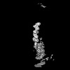
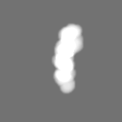
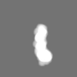
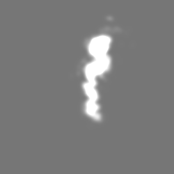
| Images |  emd_16846.png emd_16846.png | 59.9 KB | ||
| Masks | emd_16846_msk_1.map | 59.6 MB | Mask map | |
| Filedesc metadata | emd-16846.cif.gz | 5.2 KB | ||
| Others | emd_16846_additional_1.map.gzemd_16846_additional_2.map.gzemd_16846_additional_3.map.gzemd_16846_half_map_1.map.gzemd_16846_half_map_2.map.gz | 55.1 MB 3.8 MB 45.9 MB 46 MB 46 MB | ||
| Archive directory |  http://ftp.pdbj.org/pub/emdb/structures/EMD-16846ftp://ftp.pdbj.org/pub/emdb/structures/EMD-16846 http://ftp.pdbj.org/pub/emdb/structures/EMD-16846ftp://ftp.pdbj.org/pub/emdb/structures/EMD-16846 | HTTPS FTP |
-Related structure data


-Links
| EMDB pages | EMDB (EBI/PDBe) / EMDataResource |
|---|
-Map
| File | Download / File: emd_16846.map.gz / Format: CCP4 / Size: 59.6 MB / Type: IMAGE STORED AS FLOATING POINT NUMBER (4 BYTES) | ||||||||||||||||||||||||||||||||||||
|---|---|---|---|---|---|---|---|---|---|---|---|---|---|---|---|---|---|---|---|---|---|---|---|---|---|---|---|---|---|---|---|---|---|---|---|---|---|
| Annotation | Main map, DeepEMhancer post-processed | ||||||||||||||||||||||||||||||||||||
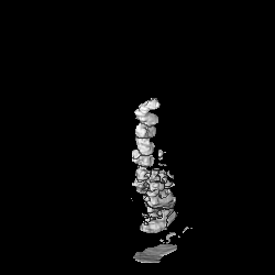
| Projections & slices | Image control
Images are generated by Spider. | ||||||||||||||||||||||||||||||||||||
| Voxel size | X=Y=Z: 2.13 Å | ||||||||||||||||||||||||||||||||||||
| Density |
| ||||||||||||||||||||||||||||||||||||
| Symmetry | Space group: 1 | ||||||||||||||||||||||||||||||||||||
| Details | EMDB XML:
|
 Z (Sec.)
 Y (Row.)
 X (Col.)


-Supplemental data
-Mask #1
| File | emd_16846_msk_1.map | ||||||||||||
|---|---|---|---|---|---|---|---|---|---|---|---|---|---|
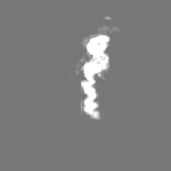
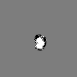
| Projections & Slices |
| ||||||||||||
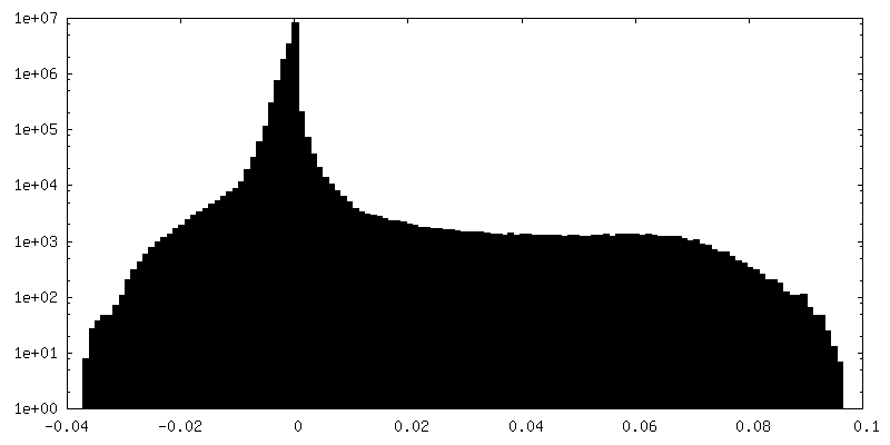
| Density Histograms |


-Additional map: Gaussian filtered map (SD 5)
| File | emd_16846_additional_1.map | ||||||||||||
|---|---|---|---|---|---|---|---|---|---|---|---|---|---|
| Annotation | Gaussian filtered map (SD 5) | ||||||||||||
| Projections & Slices |
| ||||||||||||
| Density Histograms |


-Additional map: Relion 3.1 masked post-processed map
| File | emd_16846_additional_2.map | ||||||||||||
|---|---|---|---|---|---|---|---|---|---|---|---|---|---|
| Annotation | Relion 3.1 masked post-processed map | ||||||||||||
| Projections & Slices |
| ||||||||||||
| Density Histograms |


-Additional map: Raw 3D refinement map
| File | emd_16846_additional_3.map | ||||||||||||
|---|---|---|---|---|---|---|---|---|---|---|---|---|---|
| Annotation | Raw 3D refinement map | ||||||||||||
| Projections & Slices |
| ||||||||||||
| Density Histograms |


-Half map: Half map 1
| File | emd_16846_half_map_1.map | ||||||||||||
|---|---|---|---|---|---|---|---|---|---|---|---|---|---|
| Annotation | Half map 1 | ||||||||||||
| Projections & Slices |
| ||||||||||||
| Density Histograms |


-Half map: Half map 2
| File | emd_16846_half_map_2.map | ||||||||||||
|---|---|---|---|---|---|---|---|---|---|---|---|---|---|
| Annotation | Half map 2 | ||||||||||||
| Projections & Slices |
| ||||||||||||
| Density Histograms |


- Sample components
-Entire : Myosin-5a bound to F-actin
| Entire | Name: Myosin-5a bound to F-actin |
|---|---|
| Components |
|
-Supramolecule #1: Myosin-5a bound to F-actin
| Supramolecule | Name: Myosin-5a bound to F-actin / type: complex / ID: 1 / Parent: 0 |
|---|---|
| Molecular weight | Theoretical: 20 KDa |
-Supramolecule #2: Actin, alpha skeletal muscle
| Supramolecule | Name: Actin, alpha skeletal muscle / type: complex / ID: 2 / Parent: 1 |
|---|---|
| Source (natural) | Organism: |
-Supramolecule #3: Unconventional myosin-Va bound to Calmodulin-1
| Supramolecule | Name: Unconventional myosin-Va bound to Calmodulin-1 / type: complex / ID: 3 / Parent: 1 |
|---|---|
| Source (natural) | Organism: |
-Supramolecule #4: Calmodulin-1
| Supramolecule | Name: Calmodulin-1 / type: complex / ID: 4 / Parent: 3 / Details: 1 calmodulin bound to each IQ domain, 6 in total. |
|---|---|
| Source (natural) | Organism: |
-Experimental details
-Structure determination
| Method | cryo EM |
|---|---|
 Processing Processing | single particle reconstruction |
| Aggregation state | helical array |
-Sample preparation
| Buffer | pH: 7 Component:
| |||||||||||||||
|---|---|---|---|---|---|---|---|---|---|---|---|---|---|---|---|---|
| Grid | Model: Quantifoil R2/2 / Material: COPPER / Mesh: 300 / Support film - Material: CARBON / Support film - topology: HOLEY / Pretreatment - Type: GLOW DISCHARGE / Pretreatment - Time: 30 sec. / Pretreatment - Atmosphere: AMYLAMINE | |||||||||||||||
| Vitrification | Cryogen name: ETHANE / Chamber humidity: 80 % / Chamber temperature: 281.15 K / Instrument: FEI VITROBOT MARK IV | |||||||||||||||
| Details | 5.26 mg/mL Actin, alpha skeletal muscle 266 mg/mL FlAG-tagged unconventional myosin-Va (residues 1-907) 84.2 mg/mL Calmodulin-1 |
- Electron microscopy
| Microscope | TFS KRIOS |
|---|---|
| Image recording | Film or detector model: FEI FALCON III (4k x 4k) / Detector mode: INTEGRATING / Number grids imaged: 1 / Number real images: 4285 / Average exposure time: 1.5 sec. / Average electron dose: 63.13 e/Å2 |
| Electron beam | Acceleration voltage: 300 kV / Electron source:  FIELD EMISSION GUN FIELD EMISSION GUN |
| Electron optics | C2 aperture diameter: 50.0 µm / Illumination mode: FLOOD BEAM / Imaging mode: BRIGHT FIELD / Cs: 2.7 mm / Nominal defocus max: 3.6 µm / Nominal defocus min: 1.8 µm / Nominal magnification: 75000 |
| Sample stage | Specimen holder model: FEI TITAN KRIOS AUTOGRID HOLDER / Cooling holder cryogen: NITROGEN |
| Experimental equipment |  Model: Titan Krios / Image courtesy: FEI Company |

