 Movie
Movie Controller


+ Open data

- Basic information
| Entry | Database: PDB / ID: 8kbm | ||||||
|---|---|---|---|---|---|---|---|
| Title | Structure of AcrIIA7 complexed with cGG | ||||||
 Components Components | Inhibitor of Type II CRISPR-Cas system | ||||||
 Keywords Keywords | VIRAL PROTEIN | ||||||
| Function / homology | Protein of unknown function DUF2829 / Thoeris anti-defense 2-like domain / nucleotide binding / Chem-C2E / Inhibitor of Type II CRISPR-Cas system Function and homology information Function and homology information | ||||||
| Biological species | metagenome (others) | ||||||
| Method |  X-RAY DIFFRACTION / SYNCHROTRON / SAD / Resolution: 1.38 Å X-RAY DIFFRACTION / SYNCHROTRON / SAD / Resolution: 1.38 Å | ||||||
 Authors Authors | Xiao, Y. / Feng, Y. | ||||||
| Funding support |  China, 1items China, 1items
| ||||||
 Citation Citation | Journal: Nature / Year: 2024 Title: Single phage proteins sequester signals from TIR and cGAS-like enzymes. Authors: Li, D. / Xiao, Y. / Fedorova, I. / Xiong, W. / Wang, Y. / Liu, X. / Huiting, E. / Ren, J. / Gao, Z. / Zhao, X. / Cao, X. / Zhang, Y. / Bondy-Denomy, J. / Feng, Y. | ||||||
| History |
|
- Structure visualization
| Structure viewer | Molecule: MolmilJmol/JSmol |
|---|
- Downloads & links
-Download
| PDBx/mmCIF format | 8kbm.cif.gz | 65.8 KB | Display | PDBx/mmCIF format |
|---|---|---|---|---|
| PDB format | pdb8kbm.ent.gz | 47.3 KB | Display | PDB format |
| PDBx/mmJSON format | 8kbm.json.gz | Tree view | PDBx/mmJSON format | |
| Others |  Other downloads Other downloads |
-Validation report
| Arichive directory | https://data.pdbj.org/pub/pdb/validation_reports/kb/8kbmftp://data.pdbj.org/pub/pdb/validation_reports/kb/8kbm | HTTPS FTP |
|---|
-Related structure data
| Related structure data | 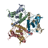 8kbbC  8kbcC 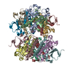 8kbdC 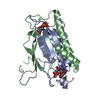 8kbeC 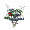 8kbfC 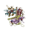 8kbgC 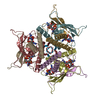 8kbhC 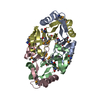 8kbiC 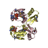 8kbjC 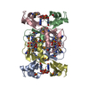 8kbkC 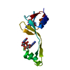 8kblC 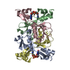 8wjcC 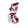 8wjdC 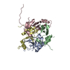 8wjeC C: citing same article ( |
|---|---|
| Similar structure data |
-Links
 PDBj
PDBj
- Assembly
| Deposited unit | 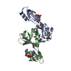
| ||||||||
|---|---|---|---|---|---|---|---|---|---|
| 1 | 
| ||||||||
| Unit cell |
| ||||||||
| Components on special symmetry positions |
|
-Components
| #1: Protein | Mass: 12016.822 Da / Num. of mol.: 2 Source method: isolated from a genetically manipulated source Source: (gene. exp.) metagenome (others) / Gene: acrIIA7 / Production host:  #2: Chemical |   Mass: 690.411 Da / Num. of mol.: 2 / Source method: obtained synthetically / Formula: C20H24N10O14P2 / Feature type: SUBJECT OF INVESTIGATION Mass: 690.411 Da / Num. of mol.: 2 / Source method: obtained synthetically / Formula: C20H24N10O14P2 / Feature type: SUBJECT OF INVESTIGATION#3: Water | ChemComp-HOH / |  Mass: 18.015 Da / Num. of mol.: 265 / Source method: isolated from a natural source / Formula: H2O Mass: 18.015 Da / Num. of mol.: 265 / Source method: isolated from a natural source / Formula: H2OHas ligand of interest | Y | Has protein modification | Y | |
|---|
-Experimental details
-Experiment
| Experiment | Method: X-RAY DIFFRACTION / Number of used crystals: 1 |
|---|
- Sample preparation
| Crystal | Density Matthews: 2.53 Å3/Da / Density % sol: 51.39 % |
|---|---|
| Crystal grow | Temperature: 291 K / Method: vapor diffusion, hanging drop Details: 1.0 M Lithium chloride, 20% w/v Polyethylene glycol 6000 |
-Data collection
| Diffraction | Mean temperature: 100 K / Serial crystal experiment: N |
|---|---|
| Diffraction source | Source: SYNCHROTRON / Site: SSRF / Beamline: BL19U1 / Wavelength: 0.979 Å |
| Detector | Type: DECTRIS PILATUS 6M / Detector: PIXEL / Date: Dec 23, 2019 |
| Radiation | Protocol: SINGLE WAVELENGTH / Monochromatic (M) / Laue (L): M / Scattering type: x-ray |
| Radiation wavelength | Wavelength: 0.979 Å / Relative weight: 1 |
| Reflection | Resolution: 1.38→19.77 Å / Num. obs: 97761 / % possible obs: 97.9 % / Redundancy: 26.4 % / Rrim(I) all: 0.109 / Net I/σ(I): 15 |
| Reflection shell | Resolution: 1.38→1.45 Å / Mean I/σ(I) obs: 2 / Num. unique obs: 4952 |
- Processing
| Software |
| ||||||||||||||||||||||||||||||||||||||||||||||||||||||||||||||||||||||||||||||||||||||||||||||||||||||||||||||||||||||||||||||||||||||||||||||||||||||||||||||||||||||||
|---|---|---|---|---|---|---|---|---|---|---|---|---|---|---|---|---|---|---|---|---|---|---|---|---|---|---|---|---|---|---|---|---|---|---|---|---|---|---|---|---|---|---|---|---|---|---|---|---|---|---|---|---|---|---|---|---|---|---|---|---|---|---|---|---|---|---|---|---|---|---|---|---|---|---|---|---|---|---|---|---|---|---|---|---|---|---|---|---|---|---|---|---|---|---|---|---|---|---|---|---|---|---|---|---|---|---|---|---|---|---|---|---|---|---|---|---|---|---|---|---|---|---|---|---|---|---|---|---|---|---|---|---|---|---|---|---|---|---|---|---|---|---|---|---|---|---|---|---|---|---|---|---|---|---|---|---|---|---|---|---|---|---|---|---|---|---|---|---|---|
| Refinement | Method to determine structure: SAD / Resolution: 1.38→19.77 Å / SU ML: 0.2 / Cross valid method: FREE R-VALUE / σ(F): 1.36 / Phase error: 28.69 / Stereochemistry target values: ML
| ||||||||||||||||||||||||||||||||||||||||||||||||||||||||||||||||||||||||||||||||||||||||||||||||||||||||||||||||||||||||||||||||||||||||||||||||||||||||||||||||||||||||
| Solvent computation | Shrinkage radii: 0.9 Å / VDW probe radii: 1.11 Å / Solvent model: FLAT BULK SOLVENT MODEL | ||||||||||||||||||||||||||||||||||||||||||||||||||||||||||||||||||||||||||||||||||||||||||||||||||||||||||||||||||||||||||||||||||||||||||||||||||||||||||||||||||||||||
| Refinement step | Cycle: LAST / Resolution: 1.38→19.77 Å
| ||||||||||||||||||||||||||||||||||||||||||||||||||||||||||||||||||||||||||||||||||||||||||||||||||||||||||||||||||||||||||||||||||||||||||||||||||||||||||||||||||||||||
| Refine LS restraints |
| ||||||||||||||||||||||||||||||||||||||||||||||||||||||||||||||||||||||||||||||||||||||||||||||||||||||||||||||||||||||||||||||||||||||||||||||||||||||||||||||||||||||||
| LS refinement shell |
|
