 Movie
Movie Controller

[English] 日本語
 Yorodumi
- PDB-8ka0: Crystal structure of Vibrio vulnificus RID-dependent transforming... -
+ Open data

- Basic information
| Entry | Database: PDB / ID: 8ka0 | |||||||||
|---|---|---|---|---|---|---|---|---|---|---|
| Title | Crystal structure of Vibrio vulnificus RID-dependent transforming NADase domain (RDTND)/calmodulin-binding domain of Rho inactivation domain (RID-CBD) complexed with Ca2+-bound calmodulin and a nicotinamide adenine dinucleotide (NAD+) | |||||||||
 Components Components |
| |||||||||
 Keywords Keywords | TOXIN / MARTX toxin / RDTND-RID / NADase / CaM / NAD+ | |||||||||
| Function / homology |  Function and homology information Function and homology informationLigases; Forming carbon-nitrogen bonds; Acid-amino-acid ligases (peptide synthases) / transporter inhibitor activity / host cell cytosol / acyltransferase activity / negative regulation of ryanodine-sensitive calcium-release channel activity / ligase activity / negative regulation of calcium ion export across plasma membrane / presynaptic endocytosis / calcineurin-mediated signaling / regulation of cell communication by electrical coupling involved in cardiac conduction ...Ligases; Forming carbon-nitrogen bonds; Acid-amino-acid ligases (peptide synthases) / transporter inhibitor activity / host cell cytosol / acyltransferase activity / negative regulation of ryanodine-sensitive calcium-release channel activity / ligase activity / negative regulation of calcium ion export across plasma membrane / presynaptic endocytosis / calcineurin-mediated signaling / regulation of cell communication by electrical coupling involved in cardiac conduction / adenylate cyclase binding / protein phosphatase activator activity / detection of calcium ion / regulation of cardiac muscle contraction / catalytic complex / calcium channel inhibitor activity / presynaptic cytosol / regulation of release of sequestered calcium ion into cytosol by sarcoplasmic reticulum / titin binding / regulation of cardiac muscle contraction by regulation of the release of sequestered calcium ion / regulation of calcium-mediated signaling / cysteine-type peptidase activity / voltage-gated potassium channel complex / Transferases; Acyltransferases; Transferring groups other than aminoacyl groups / calcium channel complex / substantia nigra development / regulation of heart rate / calyx of Held / adenylate cyclase activator activity / protein serine/threonine kinase activator activity / regulation of cytokinesis / spindle microtubule / sarcomere / calcium channel regulator activity / response to calcium ion / G2/M transition of mitotic cell cycle / spindle pole / calcium-dependent protein binding / long-term synaptic potentiation / myelin sheath / toxin activity / synaptic vesicle membrane / sperm midpiece / vesicle / transmembrane transporter binding / Hydrolases; Acting on peptide bonds (peptidases); Cysteine endopeptidases / G protein-coupled receptor signaling pathway / calcium ion binding / centrosome / lipid binding / protein kinase binding / host cell plasma membrane / protein-containing complex / proteolysis / extracellular region / membrane / metal ion binding / nucleus / plasma membrane / cytoplasm Similarity search - Function | |||||||||
| Biological species |  Vibrio vulnificus (bacteria) Vibrio vulnificus (bacteria) Homo sapiens (human) Homo sapiens (human) | |||||||||
| Method |  X-RAY DIFFRACTION / SYNCHROTRON / MOLECULAR REPLACEMENT / Resolution: 2.35 Å X-RAY DIFFRACTION / SYNCHROTRON / MOLECULAR REPLACEMENT / Resolution: 2.35 Å | |||||||||
 Authors Authors | Lee, Y. / Choi, S. / Hwang, J. / Kim, M.H. | |||||||||
| Funding support |  Korea, Republic Of, 2items Korea, Republic Of, 2items
| |||||||||
 Citation Citation | Journal: Nat Commun / Year: 2024 Title: Dissemination of pathogenic bacteria is reinforced by a MARTX toxin effector duet. Authors: Sanghyeon Choi / Youngjin Lee / Shinhye Park / Song Yee Jang / Jongbin Park / Do Won Oh / Su-Man Kim / Tae-Hwan Kim / Ga Seul Lee / Changyi Cho / Byoung Sik Kim / Donghan Lee / Eun-Hee Kim / ...Authors: Sanghyeon Choi / Youngjin Lee / Shinhye Park / Song Yee Jang / Jongbin Park / Do Won Oh / Su-Man Kim / Tae-Hwan Kim / Ga Seul Lee / Changyi Cho / Byoung Sik Kim / Donghan Lee / Eun-Hee Kim / Hae-Kap Cheong / Jeong Hee Moon / Ji-Joon Song / Jungwon Hwang / Myung Hee Kim / Abstract: Multiple bacterial genera take advantage of the multifunctional autoprocessing repeats-in-toxin (MARTX) toxin to invade host cells. Secretion of the MARTX toxin by Vibrio vulnificus, a deadly ...Multiple bacterial genera take advantage of the multifunctional autoprocessing repeats-in-toxin (MARTX) toxin to invade host cells. Secretion of the MARTX toxin by Vibrio vulnificus, a deadly opportunistic pathogen that causes primary septicemia, the precursor of sepsis, is a major driver of infection; however, the molecular mechanism via which the toxin contributes to septicemia remains unclear. Here, we report the crystal and cryo-electron microscopy (EM) structures of a toxin effector duet comprising the domain of unknown function in the first position (DUF1)/Rho inactivation domain (RID) complexed with human targets. These structures reveal how the duet is used by bacteria as a potent weapon. The data show that DUF1 acts as a RID-dependent transforming NADase domain (RDTND) that disrupts NAD homeostasis by hijacking calmodulin. The cryo-EM structure of the RDTND-RID duet complexed with calmodulin and Rac1, together with immunological analyses in vitro and in mice, provide mechanistic insight into how V. vulnificus uses the duet to suppress ROS generation by depleting NAD(P) and modifying Rac1 in a mutually-reinforcing manner that ultimately paralyzes first line immune responses, promotes dissemination of invaders, and induces sepsis. These data may allow development of tools or strategies to combat MARTX toxin-related human diseases. | |||||||||
| History |
|
- Structure visualization
| Structure viewer | Molecule: MolmilJmol/JSmol |
|---|
- Downloads & links
-Download
| PDBx/mmCIF format | 8ka0.cif.gz | 458.7 KB | Display | PDBx/mmCIF format |
|---|---|---|---|---|
| PDB format | pdb8ka0.ent.gz | 374.5 KB | Display | PDB format |
| PDBx/mmJSON format | 8ka0.json.gz | Tree view | PDBx/mmJSON format | |
| Others |  Other downloads Other downloads |
-Validation report
| Arichive directory | https://data.pdbj.org/pub/pdb/validation_reports/ka/8ka0ftp://data.pdbj.org/pub/pdb/validation_reports/ka/8ka0 | HTTPS FTP |
|---|
-Related structure data
| Related structure data | 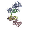 8k9zSC 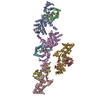 8ka1C 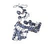 8ka2C S: Starting model for refinement C: citing same article ( |
|---|---|
| Similar structure data |
-Links
 PDBj
PDBj


- Assembly
| Deposited unit | 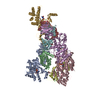
| ||||||||
|---|---|---|---|---|---|---|---|---|---|
| 1 | 
| ||||||||
| 2 | 
| ||||||||
| 3 | 
| ||||||||
| 4 | 
| ||||||||
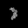
| Unit cell |
|
-Components
-Protein , 2 types, 8 molecules ACEGBDFH
| #1: Protein | Mass: 47092.160 Da / Num. of mol.: 4 / Fragment: DUF1-RIDcbd (residues, 1959-2374) / Mutation: E2186Q Source method: isolated from a genetically manipulated source Details: NCBI accession ID: WP_015728045.1 / Source: (gene. exp.) Vibrio vulnificus (bacteria) / Gene: CRN52_02910 / Production host: References: UniProt: A0A2S3R7M0, Hydrolases; Acting on peptide bonds (peptidases); Cysteine endopeptidases #2: Protein | Mass: 16979.691 Da / Num. of mol.: 4 Source method: isolated from a genetically manipulated source Details: N-terminal additional vector sequences, GA / Source: (gene. exp.) Homo sapiens (human) / Gene: CALM2, CAM2, CAMB / Production host: |
|---|
-Non-polymers , 4 types, 632 molecules 


| #3: Chemical | ChemComp-NAD /  Mass: 663.425 Da / Num. of mol.: 4 / Source method: isolated from a natural source / Formula: C21H27N7O14P2 / Feature type: SUBJECT OF INVESTIGATION / Comment: NAD*YM Mass: 663.425 Da / Num. of mol.: 4 / Source method: isolated from a natural source / Formula: C21H27N7O14P2 / Feature type: SUBJECT OF INVESTIGATION / Comment: NAD*YM#4: Chemical | ChemComp-CA /  Mass: 40.078 Da / Num. of mol.: 8 / Source method: obtained synthetically / Formula: Ca / Feature type: SUBJECT OF INVESTIGATION Mass: 40.078 Da / Num. of mol.: 8 / Source method: obtained synthetically / Formula: Ca / Feature type: SUBJECT OF INVESTIGATION#5: Chemical | ChemComp-GOL /  Mass: 92.094 Da / Num. of mol.: 4 / Source method: obtained synthetically / Formula: C3H8O3 Mass: 92.094 Da / Num. of mol.: 4 / Source method: obtained synthetically / Formula: C3H8O3#6: Water | ChemComp-HOH / | Mass: 18.015 Da / Num. of mol.: 616 / Source method: isolated from a natural source / Formula: H2O |
|---|
-Details
| Has ligand of interest | Y |
|---|
-Experimental details
-Experiment
| Experiment | Method: X-RAY DIFFRACTION / Number of used crystals: 1 |
|---|
- Sample preparation
| Crystal | Density Matthews: 2.38 Å3/Da / Density % sol: 48.25 % |
|---|---|
| Crystal grow | Temperature: 293 K / Method: vapor diffusion, sitting drop / pH: 8.5 Details: 0.1 M Tris-HCl (pH 8.5), 32.5% (w/v) PEG 4000 and 0.15 M sodium acetate. 10 mM NAD+ soaking |
-Data collection
| Diffraction | Mean temperature: 100 K / Serial crystal experiment: N |
|---|---|
| Diffraction source | Source: SYNCHROTRON / Site: PAL/PLS / Beamline: 5C (4A) / Wavelength: 1 Å |
| Detector | Type: DECTRIS EIGER X 9M / Detector: PIXEL / Date: Mar 24, 2022 |
| Radiation | Protocol: SINGLE WAVELENGTH / Monochromatic (M) / Laue (L): M / Scattering type: x-ray |
| Radiation wavelength | Wavelength: 1 Å / Relative weight: 1 |
| Reflection | Resolution: 2.35→29.62 Å / Num. obs: 98734 / % possible obs: 99.8 % / Redundancy: 6.9 % / CC1/2: 0.999 / Net I/σ(I): 11.6 |
| Reflection shell | Resolution: 2.35→2.39 Å / Num. unique obs: 4831 / CC1/2: 0.675 / % possible all: 99.9 |
- Processing
| Software |
| ||||||||||||||||||||||||||||||||||||||||||||||||||||||||||||||||||||||||||||||||||||||||||||||||||||||||||||||||||||||||||||||||||||||||||||||||||||||||||||||||||||||||||||||||||||||
|---|---|---|---|---|---|---|---|---|---|---|---|---|---|---|---|---|---|---|---|---|---|---|---|---|---|---|---|---|---|---|---|---|---|---|---|---|---|---|---|---|---|---|---|---|---|---|---|---|---|---|---|---|---|---|---|---|---|---|---|---|---|---|---|---|---|---|---|---|---|---|---|---|---|---|---|---|---|---|---|---|---|---|---|---|---|---|---|---|---|---|---|---|---|---|---|---|---|---|---|---|---|---|---|---|---|---|---|---|---|---|---|---|---|---|---|---|---|---|---|---|---|---|---|---|---|---|---|---|---|---|---|---|---|---|---|---|---|---|---|---|---|---|---|---|---|---|---|---|---|---|---|---|---|---|---|---|---|---|---|---|---|---|---|---|---|---|---|---|---|---|---|---|---|---|---|---|---|---|---|---|---|---|---|
| Refinement | Method to determine structure: MOLECULAR REPLACEMENT Starting model: 8K9Z Resolution: 2.35→29.62 Å / Cor.coef. Fo:Fc: 0.952 / Cor.coef. Fo:Fc free: 0.924 / Cross valid method: THROUGHOUT / ESU R: 0.494 / ESU R Free: 0.279 / Stereochemistry target values: MAXIMUM LIKELIHOOD / Details: HYDROGENS HAVE BEEN ADDED IN THE RIDING POSITIONS
| ||||||||||||||||||||||||||||||||||||||||||||||||||||||||||||||||||||||||||||||||||||||||||||||||||||||||||||||||||||||||||||||||||||||||||||||||||||||||||||||||||||||||||||||||||||||
| Solvent computation | Ion probe radii: 0.8 Å / Shrinkage radii: 0.8 Å / VDW probe radii: 1.2 Å / Solvent model: MASK | ||||||||||||||||||||||||||||||||||||||||||||||||||||||||||||||||||||||||||||||||||||||||||||||||||||||||||||||||||||||||||||||||||||||||||||||||||||||||||||||||||||||||||||||||||||||
| Displacement parameters | Biso mean: 55.8 Å2
| ||||||||||||||||||||||||||||||||||||||||||||||||||||||||||||||||||||||||||||||||||||||||||||||||||||||||||||||||||||||||||||||||||||||||||||||||||||||||||||||||||||||||||||||||||||||
| Refinement step | Cycle: LAST / Resolution: 2.35→29.62 Å
| ||||||||||||||||||||||||||||||||||||||||||||||||||||||||||||||||||||||||||||||||||||||||||||||||||||||||||||||||||||||||||||||||||||||||||||||||||||||||||||||||||||||||||||||||||||||
| Refine LS restraints |
|