 Movie
Movie Controller


+ Open data

- Basic information
| Entry | Database: PDB / ID: 8k7r | |||||||||||||||||||||||||||||||||||||||
|---|---|---|---|---|---|---|---|---|---|---|---|---|---|---|---|---|---|---|---|---|---|---|---|---|---|---|---|---|---|---|---|---|---|---|---|---|---|---|---|---|
| Title | Human Fc epsilon RI in complex with hIgE Fc (TMD disordered) | |||||||||||||||||||||||||||||||||||||||
 Components Components |
| |||||||||||||||||||||||||||||||||||||||
 Keywords Keywords | IMMUNE SYSTEM / IgE / high-affinity IgE receptor / allergy | |||||||||||||||||||||||||||||||||||||||
| Function / homology |  Function and homology information Function and homology informationhigh-affinity IgE receptor activity / type I hypersensitivity / eosinophil degranulation / IgE binding / Fc epsilon receptor (FCERI) signaling / type 2 immune response / mast cell degranulation / immunoglobulin mediated immune response / Role of LAT2/NTAL/LAB on calcium mobilization / FCERI mediated Ca+2 mobilization ...high-affinity IgE receptor activity / type I hypersensitivity / eosinophil degranulation / IgE binding / Fc epsilon receptor (FCERI) signaling / type 2 immune response / mast cell degranulation / immunoglobulin mediated immune response / Role of LAT2/NTAL/LAB on calcium mobilization / FCERI mediated Ca+2 mobilization / FCERI mediated MAPK activation / FCERI mediated NF-kB activation / cell surface receptor signaling pathway / external side of plasma membrane / cell surface / plasma membrane Similarity search - Function | |||||||||||||||||||||||||||||||||||||||
| Biological species |  Homo sapiens (human) Homo sapiens (human) | |||||||||||||||||||||||||||||||||||||||
| Method | ELECTRON MICROSCOPY / single particle reconstruction / cryo EM / Resolution: 3.56 Å | |||||||||||||||||||||||||||||||||||||||
 Authors Authors | Zhang, Z. / Yui, M. / Ohto, U. / Shimizu, T. | |||||||||||||||||||||||||||||||||||||||
| Funding support | 1items
| |||||||||||||||||||||||||||||||||||||||
 Citation Citation | Journal: Sci Signal / Year: 2024 Title: Architecture of the high-affinity immunoglobulin E receptor. Authors: Zhikuan Zhang / Moeko Yui / Umeharu Ohto / Toshiyuki Shimizu /  Abstract: The high-affinity immunoglobulin E (IgE) receptor (FcεRI) drives type I hypersensitivity in response to allergen-specific IgE. FcεRI is a multimeric complex typically composed of one α, one β, ...The high-affinity immunoglobulin E (IgE) receptor (FcεRI) drives type I hypersensitivity in response to allergen-specific IgE. FcεRI is a multimeric complex typically composed of one α, one β, and two disulfide-linked γ subunits. The α subunit binds to the fragment crystallizable (Fc) region of IgE (Fcε), whereas the β and γ subunits mediate signaling through their intracellular immunoreceptor tyrosine-based activation motifs (ITAMs). Here, we report cryo-electron microscopy (cryo-EM) structures of the apo state of FcεRI and of FcεRI bound to Fcε. At the transmembrane domain (TMD), the α and γ subunits associate to form a tightly packed, three-helix bundle (αγ bundle) with pseudo-threefold symmetry through extensive hydrophobic and polar interactions. The αγ bundle further assembles with the β subunit to complete the TMD, from which multiple ITAMs might extend into the cytoplasm for downstream signaling. The apo mouse FcεRI essentially forms an identical structure to that of the Fcε-bound sensitized form, suggesting that the binding of Fcε to FcεRI does not alter the overall conformation of the receptor. Furthermore, the juxtamembrane interaction between the extracellular domains (ECDs) of mouse FcεRIα and FcεRIβ is not observed between their human counterparts, which implies potential species-specific differences in receptor stability and activation. Our findings provide a framework for understanding the general structural principles underlying Fc receptor assembly, the signaling mechanism underlying type I hypersensitivity, and the design of efficient antiallergic therapeutics. | |||||||||||||||||||||||||||||||||||||||
| History |
|
- Structure visualization
| Structure viewer | Molecule: MolmilJmol/JSmol |
|---|
- Downloads & links
-Download
| PDBx/mmCIF format | 8k7r.cif.gz | 160.9 KB | Display | PDBx/mmCIF format |
|---|---|---|---|---|
| PDB format | pdb8k7r.ent.gz | 123.8 KB | Display | PDB format |
| PDBx/mmJSON format | 8k7r.json.gz | Tree view | PDBx/mmJSON format | |
| Others |  Other downloads Other downloads |
-Validation report
| Arichive directory | https://data.pdbj.org/pub/pdb/validation_reports/k7/8k7rftp://data.pdbj.org/pub/pdb/validation_reports/k7/8k7r | HTTPS FTP |
|---|
-Related structure data
| Related structure data |  36939MC  8k7sC  8k7tC 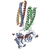 8yrjC M: map data used to model this data C: citing same article ( |
|---|---|
| Similar structure data |


-Links
 PDBj
PDBj


- Assembly
| Deposited unit | 
|
|---|---|
| 1 |
|
-Components
-Protein , 2 types, 3 molecules AEF
| #1: Protein | Mass: 32578.768 Da / Num. of mol.: 1 Source method: isolated from a genetically manipulated source Source: (gene. exp.) Homo sapiens (human) / Gene: FCER1A / Production host: Homo sapiens (human) / References: UniProt: P12319 |
|---|---|
| #2: Protein | Mass: 39236.145 Da / Num. of mol.: 2 Source method: isolated from a genetically manipulated source Source: (gene. exp.) Homo sapiens (human) / Production host: Homo sapiens (human) |
-Sugars , 5 types, 7 molecules 
| #3: Polysaccharide | 2-acetamido-2-deoxy-beta-D-glucopyranose-(1-4)-2-acetamido-2-deoxy-beta-D-glucopyranose Source method: isolated from a genetically manipulated source | ||
|---|---|---|---|
| #4: Polysaccharide | 2-acetamido-2-deoxy-beta-D-glucopyranose-(1-4)-[alpha-L-fucopyranose-(1-6)]2-acetamido-2-deoxy-beta- ...2-acetamido-2-deoxy-beta-D-glucopyranose-(1-4)-[alpha-L-fucopyranose-(1-6)]2-acetamido-2-deoxy-beta-D-glucopyranose Source method: isolated from a genetically manipulated source | ||
| #5: Polysaccharide | alpha-D-mannopyranose-(1-3)-beta-D-mannopyranose-(1-4)-2-acetamido-2-deoxy-beta-D-glucopyranose-(1- ...alpha-D-mannopyranose-(1-3)-beta-D-mannopyranose-(1-4)-2-acetamido-2-deoxy-beta-D-glucopyranose-(1-4)-2-acetamido-2-deoxy-beta-D-glucopyranose Source method: isolated from a genetically manipulated source | ||
| #6: Polysaccharide | Source method: isolated from a genetically manipulated source #7: Sugar |  Type: D-saccharide, beta linking / Mass: 221.208 Da / Num. of mol.: 2 / Source method: obtained synthetically / Formula: C8H15NO6 Type: D-saccharide, beta linking / Mass: 221.208 Da / Num. of mol.: 2 / Source method: obtained synthetically / Formula: C8H15NO6 |
-Details
| Has ligand of interest | N |
|---|---|
| Has protein modification | Y |
-Experimental details
-Experiment
| Experiment | Method: ELECTRON MICROSCOPY |
|---|---|
| EM experiment | Aggregation state: PARTICLE / 3D reconstruction method: single particle reconstruction |
- Sample preparation
| Component | Name: Human Fc epsilon RI in complex with hIgE Fc / Type: COMPLEX / Entity ID: #1-#2 / Source: RECOMBINANT |
|---|---|
| Molecular weight | Experimental value: NO |
| Source (natural) | Organism: Homo sapiens (human) |
| Source (recombinant) | Organism: Homo sapiens (human) |
| Buffer solution | pH: 7.7 |
| Specimen | Embedding applied: NO / Shadowing applied: NO / Staining applied: NO / Vitrification applied: YES |
| Vitrification | Cryogen name: ETHANE |
- Electron microscopy imaging
| Experimental equipment |  Model: Titan Krios / Image courtesy: FEI Company |
|---|---|
| Microscopy | Model: TFS KRIOS |
| Electron gun | Electron source:  FIELD EMISSION GUN / Accelerating voltage: 300 kV / Illumination mode: OTHER FIELD EMISSION GUN / Accelerating voltage: 300 kV / Illumination mode: OTHER |
| Electron lens | Mode: BRIGHT FIELD / Nominal defocus max: 2000 nm / Nominal defocus min: 800 nm |
| Image recording | Electron dose: 61 e/Å2 / Film or detector model: GATAN K3 BIOQUANTUM (6k x 4k) |
- Processing
| EM software | Name: PHENIX / Category: model refinement | ||||||||||||||||||||||||
|---|---|---|---|---|---|---|---|---|---|---|---|---|---|---|---|---|---|---|---|---|---|---|---|---|---|
| CTF correction | Type: NONE | ||||||||||||||||||||||||
| 3D reconstruction | Resolution: 3.56 Å / Resolution method: FSC 0.143 CUT-OFF / Num. of particles: 303726 / Symmetry type: POINT | ||||||||||||||||||||||||
| Refine LS restraints |
|
