 Movie
Movie Controller


+ Open data

- Basic information
| Entry |  | |||||||||
|---|---|---|---|---|---|---|---|---|---|---|
| Title | Human Fc epsilon RI in complex with hIgE Fc (TMD disordered) | |||||||||
 Map data Map data | Sharpened map | |||||||||
 Sample Sample |
| |||||||||
 Keywords Keywords | IgE / high-affinity IgE receptor / allergy / IMMUNE SYSTEM | |||||||||
| Function / homology |  Function and homology information Function and homology informationhigh-affinity IgE receptor activity / type I hypersensitivity / eosinophil degranulation / IgE binding / Fc epsilon receptor (FCERI) signaling / type 2 immune response / mast cell degranulation / immunoglobulin mediated immune response / Role of LAT2/NTAL/LAB on calcium mobilization / FCERI mediated Ca+2 mobilization ...high-affinity IgE receptor activity / type I hypersensitivity / eosinophil degranulation / IgE binding / Fc epsilon receptor (FCERI) signaling / type 2 immune response / mast cell degranulation / immunoglobulin mediated immune response / Role of LAT2/NTAL/LAB on calcium mobilization / FCERI mediated Ca+2 mobilization / FCERI mediated MAPK activation / FCERI mediated NF-kB activation / cell surface receptor signaling pathway / external side of plasma membrane / cell surface / plasma membrane Similarity search - Function | |||||||||
| Biological species |  Homo sapiens (human) Homo sapiens (human) | |||||||||
| Method | single particle reconstruction / cryo EM / Resolution: 3.56 Å | |||||||||
 Authors Authors | Zhang Z / Yui M / Ohto U / Shimizu T | |||||||||
| Funding support | 1 items
| |||||||||
 Citation Citation | Journal: Sci Signal / Year: 2024 Title: Architecture of the high-affinity immunoglobulin E receptor. Authors: Zhikuan Zhang / Moeko Yui / Umeharu Ohto / Toshiyuki Shimizu /  Abstract: The high-affinity immunoglobulin E (IgE) receptor (FcεRI) drives type I hypersensitivity in response to allergen-specific IgE. FcεRI is a multimeric complex typically composed of one α, one β, ...The high-affinity immunoglobulin E (IgE) receptor (FcεRI) drives type I hypersensitivity in response to allergen-specific IgE. FcεRI is a multimeric complex typically composed of one α, one β, and two disulfide-linked γ subunits. The α subunit binds to the fragment crystallizable (Fc) region of IgE (Fcε), whereas the β and γ subunits mediate signaling through their intracellular immunoreceptor tyrosine-based activation motifs (ITAMs). Here, we report cryo-electron microscopy (cryo-EM) structures of the apo state of FcεRI and of FcεRI bound to Fcε. At the transmembrane domain (TMD), the α and γ subunits associate to form a tightly packed, three-helix bundle (αγ bundle) with pseudo-threefold symmetry through extensive hydrophobic and polar interactions. The αγ bundle further assembles with the β subunit to complete the TMD, from which multiple ITAMs might extend into the cytoplasm for downstream signaling. The apo mouse FcεRI essentially forms an identical structure to that of the Fcε-bound sensitized form, suggesting that the binding of Fcε to FcεRI does not alter the overall conformation of the receptor. Furthermore, the juxtamembrane interaction between the extracellular domains (ECDs) of mouse FcεRIα and FcεRIβ is not observed between their human counterparts, which implies potential species-specific differences in receptor stability and activation. Our findings provide a framework for understanding the general structural principles underlying Fc receptor assembly, the signaling mechanism underlying type I hypersensitivity, and the design of efficient antiallergic therapeutics. | |||||||||
| History |
|
- Structure visualization
| Supplemental images |
|---|

- Downloads & links
-EMDB archive
| Map data | emd_36939.map.gz | 21 MB | EMDB map data format | |
|---|---|---|---|---|
| Header (meta data) | emd-36939-v30.xmlemd-36939.xml | 19.3 KB 19.3 KB | Display Display | EMDB header |
| Images |  emd_36939.png emd_36939.png | 78.1 KB | ||
| Filedesc metadata | emd-36939.cif.gz | 6.5 KB | ||
| Others | emd_36939_additional_1.map.gzemd_36939_half_map_1.map.gzemd_36939_half_map_2.map.gz | 11.1 MB 20.6 MB 20.6 MB | ||
| Archive directory |  http://ftp.pdbj.org/pub/emdb/structures/EMD-36939ftp://ftp.pdbj.org/pub/emdb/structures/EMD-36939 http://ftp.pdbj.org/pub/emdb/structures/EMD-36939ftp://ftp.pdbj.org/pub/emdb/structures/EMD-36939 | HTTPS FTP |
-Related structure data
| Related structure data |  8k7rMC  8k7sC  8k7tC 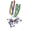 8yrjC M: atomic model generated by this map C: citing same article ( |
|---|---|
| Similar structure data |


-Links
| EMDB pages | EMDB (EBI/PDBe) / EMDataResource |
|---|---|
| Related items in Molecule of the Month |


-Map
| File | Download / File: emd_36939.map.gz / Format: CCP4 / Size: 22.2 MB / Type: IMAGE STORED AS FLOATING POINT NUMBER (4 BYTES) | ||||||||||||||||||||||||||||||||||||
|---|---|---|---|---|---|---|---|---|---|---|---|---|---|---|---|---|---|---|---|---|---|---|---|---|---|---|---|---|---|---|---|---|---|---|---|---|---|
| Annotation | Sharpened map | ||||||||||||||||||||||||||||||||||||
| Projections & slices | Image control
Images are generated by Spider. | ||||||||||||||||||||||||||||||||||||
| Voxel size | X=Y=Z: 1.38333 Å | ||||||||||||||||||||||||||||||||||||
| Density |
| ||||||||||||||||||||||||||||||||||||
| Symmetry | Space group: 1 | ||||||||||||||||||||||||||||||||||||
| Details | EMDB XML:
|
 Z (Sec.)
 Y (Row.)
 X (Col.)


-Supplemental data
-Additional map: Unsharpened map
| File | emd_36939_additional_1.map | ||||||||||||
|---|---|---|---|---|---|---|---|---|---|---|---|---|---|
| Annotation | Unsharpened map | ||||||||||||
| Projections & Slices |
| ||||||||||||
| Density Histograms |


-Half map: Half map 2
| File | emd_36939_half_map_1.map | ||||||||||||
|---|---|---|---|---|---|---|---|---|---|---|---|---|---|
| Annotation | Half map 2 | ||||||||||||
| Projections & Slices |
| ||||||||||||
| Density Histograms |


-Half map: Half map 1
| File | emd_36939_half_map_2.map | ||||||||||||
|---|---|---|---|---|---|---|---|---|---|---|---|---|---|
| Annotation | Half map 1 | ||||||||||||
| Projections & Slices |
| ||||||||||||
| Density Histograms |


- Sample components
-Entire : Human Fc epsilon RI in complex with hIgE Fc
| Entire | Name: Human Fc epsilon RI in complex with hIgE Fc |
|---|---|
| Components |
|
-Supramolecule #1: Human Fc epsilon RI in complex with hIgE Fc
| Supramolecule | Name: Human Fc epsilon RI in complex with hIgE Fc / type: complex / ID: 1 / Parent: 0 / Macromolecule list: #1-#2 |
|---|---|
| Source (natural) | Organism: Homo sapiens (human) |
-Macromolecule #1: High affinity immunoglobulin epsilon receptor subunit alpha
| Macromolecule | Name: High affinity immunoglobulin epsilon receptor subunit alpha type: protein_or_peptide / ID: 1 / Number of copies: 1 / Enantiomer: LEVO |
|---|---|
| Source (natural) | Organism: Homo sapiens (human) |
| Molecular weight | Theoretical: 32.578768 KDa |
| Recombinant expression | Organism: Homo sapiens (human) |
| Sequence | String: MAPAMESPTL LCVALLFFAP DGVLAVPQKP KVSLNPPWNR IFKGENVTLT CNGNNFFEVS STKWFHNGSL SEETNSSLNI VNAKFEDSG EYKCQHQQVN ESEPVYLEVF SDWLLLQASA EVVMEGQPLF LRCHGWRNWD VYKVIYYKDG EALKYWYENH N ISITNATV ...String: MAPAMESPTL LCVALLFFAP DGVLAVPQKP KVSLNPPWNR IFKGENVTLT CNGNNFFEVS STKWFHNGSL SEETNSSLNI VNAKFEDSG EYKCQHQQVN ESEPVYLEVF SDWLLLQASA EVVMEGQPLF LRCHGWRNWD VYKVIYYKDG EALKYWYENH N ISITNATV EDSGTYYCTG KVWQLDYESE PLNITVIKAP REKYWLQFFI PLLVVILFAV DTGLFISTQQ QVTFLLKIKR TR KGFRLLN PHPKPNPKNN ENLYFQGDYK DDDDKHHHHH HHH UniProtKB: High affinity immunoglobulin epsilon receptor subunit alpha |
-Macromolecule #2: IgE Fc
| Macromolecule | Name: IgE Fc / type: protein_or_peptide / ID: 2 / Number of copies: 2 / Enantiomer: LEVO |
|---|---|
| Source (natural) | Organism: Homo sapiens (human) |
| Molecular weight | Theoretical: 39.236145 KDa |
| Recombinant expression | Organism: Homo sapiens (human) |
| Sequence | String: METGLRWLLL VAVLKGVQCQ SVASRDFTPP TVKILQSSCD GGGHFPPTIQ LLCLVSGYTP GTIQITWLED GQVMDVDLST ASTTQEGEL ASTQSELTLS QKHWLSDRTY TCQVTYQGHT FEDSTKKCAD SNPRGVSAYL SRPSPFDLFI RKSPTITCLV V DLAPSKGT ...String: METGLRWLLL VAVLKGVQCQ SVASRDFTPP TVKILQSSCD GGGHFPPTIQ LLCLVSGYTP GTIQITWLED GQVMDVDLST ASTTQEGEL ASTQSELTLS QKHWLSDRTY TCQVTYQGHT FEDSTKKCAD SNPRGVSAYL SRPSPFDLFI RKSPTITCLV V DLAPSKGT VQLTWSRASG KPVNHSTRKE EKQRNGTLTV TSTLPVGTRD WIEGETYQCR VTHPHLPRAL MRSTTKTSGP RA APEVYAF ATPEWPGSRD KRTLACLIQN FMPEDISVQW LHNEVQLPDA RHSTTQPRKT KGSGFFVFSR LEVTRAEWEQ KDE FICRAV HEAASPSQTV QRAVSVNPHH HHHHH |
-Macromolecule #7: 2-acetamido-2-deoxy-beta-D-glucopyranose
| Macromolecule | Name: 2-acetamido-2-deoxy-beta-D-glucopyranose / type: ligand / ID: 7 / Number of copies: 2 / Formula: NAG |
|---|---|
| Molecular weight | Theoretical: 221.208 Da |
| Chemical component information |  ChemComp-NAG: |
-Experimental details
-Structure determination
| Method | cryo EM |
|---|---|
 Processing Processing | single particle reconstruction |
| Aggregation state | particle |
-Sample preparation
| Buffer | pH: 7.7 |
|---|---|
| Vitrification | Cryogen name: ETHANE |
- Electron microscopy
| Microscope | TFS KRIOS |
|---|---|
| Image recording | Film or detector model: GATAN K3 BIOQUANTUM (6k x 4k) / Average electron dose: 61.0 e/Å2 |
| Electron beam | Acceleration voltage: 300 kV / Electron source:  FIELD EMISSION GUN FIELD EMISSION GUN |
| Electron optics | Illumination mode: OTHER / Imaging mode: BRIGHT FIELD / Nominal defocus max: 2.0 µm / Nominal defocus min: 0.8 µm |
| Experimental equipment |  Model: Titan Krios / Image courtesy: FEI Company |
