 Movie
Movie Controller

[English] 日本語
 Yorodumi
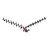
- PDB-8k5o: Cryo-EM structure of the RC-LH core comples from Halorhodospira h... -
+ Open data

- Basic information
| Entry | Database: PDB / ID: 8k5o | ||||||
|---|---|---|---|---|---|---|---|
| Title | Cryo-EM structure of the RC-LH core comples from Halorhodospira halochloris | ||||||
 Components Components |
| ||||||
 Keywords Keywords | PHOTOSYNTHESIS / RC-LH complex / HALORHODOSPIRA HALOCHLORIS | ||||||
| Function / homology |  Function and homology information Function and homology informationorganelle inner membrane / plasma membrane-derived chromatophore membrane / plasma membrane light-harvesting complex / bacteriochlorophyll binding / photosynthetic electron transport in photosystem II / photosynthesis, light reaction / endomembrane system / electron transfer activity / iron ion binding / heme binding ...organelle inner membrane / plasma membrane-derived chromatophore membrane / plasma membrane light-harvesting complex / bacteriochlorophyll binding / photosynthetic electron transport in photosystem II / photosynthesis, light reaction / endomembrane system / electron transfer activity / iron ion binding / heme binding / metal ion binding / plasma membrane Similarity search - Function | ||||||
| Biological species |  Halorhodospira halochloris (bacteria) Halorhodospira halochloris (bacteria) | ||||||
| Method | ELECTRON MICROSCOPY / single particle reconstruction / cryo EM / Resolution: 2.42 Å | ||||||
 Authors Authors | Wang, G.-L. / Qi, C.-H. / Yu, L.-J. | ||||||
| Funding support |  China, 1items China, 1items
| ||||||
 Citation Citation | Journal: J Integr Plant Biol / Year: 2024 Title: Structural insights into the unusual core photocomplex from a triply extremophilic purple bacterium, Halorhodospira halochloris. Authors: Chen-Hui Qi / Guang-Lei Wang / Fang-Fang Wang / Jie Wang / Xiang-Ping Wang / Mei-Juan Zou / Fei Ma / Michael T Madigan / Yukihiro Kimura / Zheng-Yu Wang-Otomo / Long-Jiang Yu /   Abstract: Halorhodospira (Hlr.) halochloris is a triply extremophilic phototrophic purple sulfur bacterium, as it is thermophilic, alkaliphilic, and extremely halophilic. The light-harvesting-reaction center ...Halorhodospira (Hlr.) halochloris is a triply extremophilic phototrophic purple sulfur bacterium, as it is thermophilic, alkaliphilic, and extremely halophilic. The light-harvesting-reaction center (LH1-RC) core complex of this bacterium displays an LH1-Q transition at 1,016 nm, which is the lowest-energy wavelength absorption among all known phototrophs. Here we report the cryo-EM structure of the LH1-RC at 2.42 Å resolution. The LH1 complex forms a tricyclic ring structure composed of 16 αβγ-polypeptides and one αβ-heterodimer around the RC. From the cryo-EM density map, two previously unrecognized integral membrane proteins, referred to as protein G and protein Q, were identified. Both of these proteins are single transmembrane-spanning helices located between the LH1 ring and the RC L-subunit and are absent from the LH1-RC complexes of all other purple bacteria of which the structures have been determined so far. Besides bacteriochlorophyll b molecules (B1020) located on the periplasmic side of the Hlr. halochloris membrane, there are also two arrays of bacteriochlorophyll b molecules (B800 and B820) located on the cytoplasmic side. Only a single copy of a carotenoid (lycopene) was resolved in the Hlr. halochloris LH1-α3β3 and this was positioned within the complex. The potential quinone channel should be the space between the LH1-α3β3 that accommodates the single lycopene but does not contain a γ-polypeptide, B800 and B820. Our results provide a structural explanation for the unusual Q red shift and carotenoid absorption in the Hlr. halochloris spectrum and reveal new insights into photosynthetic mechanisms employed by a species that thrives under the harshest conditions of any phototrophic microorganism known. | ||||||
| History |
|
- Structure visualization
| Structure viewer | Molecule: MolmilJmol/JSmol |
|---|
- Downloads & links
-Download
| PDBx/mmCIF format | 8k5o.cif.gz | 854.3 KB | Display | PDBx/mmCIF format |
|---|---|---|---|---|
| PDB format | pdb8k5o.ent.gz | Display | PDB format | |
| PDBx/mmJSON format | 8k5o.json.gz | Tree view | PDBx/mmJSON format | |
| Others |  Other downloads Other downloads |
-Validation report
| Arichive directory | https://data.pdbj.org/pub/pdb/validation_reports/k5/8k5oftp://data.pdbj.org/pub/pdb/validation_reports/k5/8k5o | HTTPS FTP |
|---|
-Related structure data
| Related structure data |  36907MC M: map data used to model this data C: citing same article ( |
|---|---|
| Similar structure data |
-Links
 PDBj
PDBj


- Assembly
| Deposited unit | 
|
|---|---|
| 1 |
|
-Components
-Photosynthetic reaction center ... , 2 types, 2 molecules CH
| #1: Protein | Mass: 41612.859 Da / Num. of mol.: 1 / Source method: isolated from a natural source / Source: (natural) Halorhodospira halochloris (bacteria) / References: UniProt: A0A0X8X829 |
|---|---|
| #4: Protein | Mass: 31293.768 Da / Num. of mol.: 1 / Source method: isolated from a natural source / Source: (natural) Halorhodospira halochloris (bacteria) / References: UniProt: A0A0X8X838 |
-Reaction center protein ... , 2 types, 2 molecules LM
| #2: Protein | Mass: 31115.025 Da / Num. of mol.: 1 / Source method: isolated from a natural source / Source: (natural) Halorhodospira halochloris (bacteria) / References: UniProt: A0A0X8XAH6 |
|---|---|
| #3: Protein | Mass: 36221.676 Da / Num. of mol.: 1 / Source method: isolated from a natural source / Source: (natural) Halorhodospira halochloris (bacteria) / References: UniProt: A0A0X8X847 |
-Antenna complex alpha/beta subunit domain-containing ... , 2 types, 17 molecules 4wYbkntz6FKPSVehq
| #5: Protein | Mass: 12053.332 Da / Num. of mol.: 1 / Source method: isolated from a natural source / Source: (natural) Halorhodospira halochloris (bacteria) / References: UniProt: A0A110B4Z6 |
|---|---|
| #7: Protein | Mass: 7696.815 Da / Num. of mol.: 16 / Source method: isolated from a natural source / Source: (natural) Halorhodospira halochloris (bacteria) / References: UniProt: A0A0X8XBE4 |
-Protein , 2 types, 17 molecules 3xZclou17GNQTWfir
| #6: Protein | Mass: 7562.918 Da / Num. of mol.: 1 / Source method: isolated from a natural source / Source: (natural) Halorhodospira halochloris (bacteria) / References: UniProt: A0A120MZP7 |
|---|---|
| #8: Protein | Mass: 9555.675 Da / Num. of mol.: 16 / Source method: isolated from a natural source / Source: (natural) Halorhodospira halochloris (bacteria) / References: UniProt: A0A0X8X9B2 |
-Protein/peptide , 3 types, 18 molecules vXajmsy58IORUdgp29
| #9: Protein/peptide | Mass: 3123.757 Da / Num. of mol.: 16 / Source method: isolated from a natural source / Source: (natural) Halorhodospira halochloris (bacteria)#10: Protein/peptide | | Mass: 3531.281 Da / Num. of mol.: 1 / Source method: isolated from a natural source / Source: (natural) Halorhodospira halochloris (bacteria)#11: Protein/peptide | | Mass: 3762.490 Da / Num. of mol.: 1 / Source method: isolated from a natural source / Source: (natural) Halorhodospira halochloris (bacteria) |
|---|
-Sugars , 1 types, 2 molecules 
| #25: Sugar |  Type: D-saccharide, beta linking / Mass: 292.369 Da / Num. of mol.: 2 / Source method: obtained synthetically / Formula: C14H28O6 / Comment: detergent*YM Type: D-saccharide, beta linking / Mass: 292.369 Da / Num. of mol.: 2 / Source method: obtained synthetically / Formula: C14H28O6 / Comment: detergent*YM |
|---|
-Non-polymers , 13 types, 193 molecules 


| #12: Chemical | ChemComp-HEC /  Mass: 618.503 Da / Num. of mol.: 4 / Source method: obtained synthetically / Formula: C34H34FeN4O4 / Feature type: SUBJECT OF INVESTIGATION Mass: 618.503 Da / Num. of mol.: 4 / Source method: obtained synthetically / Formula: C34H34FeN4O4 / Feature type: SUBJECT OF INVESTIGATION#13: Chemical | ChemComp-PGV / (  Mass: 749.007 Da / Num. of mol.: 50 / Source method: obtained synthetically / Formula: C40H77O10P / Comment: phospholipid*YM Mass: 749.007 Da / Num. of mol.: 50 / Source method: obtained synthetically / Formula: C40H77O10P / Comment: phospholipid*YM#14: Chemical | ChemComp-LHG / |  Mass: 722.970 Da / Num. of mol.: 1 / Source method: obtained synthetically / Formula: C38H75O10P / Comment: phospholipid*YM Mass: 722.970 Da / Num. of mol.: 1 / Source method: obtained synthetically / Formula: C38H75O10P / Comment: phospholipid*YM#15: Chemical | ChemComp-A1LZM / Mass: 907.472 Da / Num. of mol.: 54 / Source method: obtained synthetically / Formula: C55H70MgN4O6 / Feature type: SUBJECT OF INVESTIGATION #16: Chemical | Mass: 885.183 Da / Num. of mol.: 2 / Source method: obtained synthetically / Formula: C55H72N4O6 / Feature type: SUBJECT OF INVESTIGATION #17: Chemical | ChemComp-UQ8 / |  Mass: 727.109 Da / Num. of mol.: 1 / Source method: obtained synthetically / Formula: C49H74O4 / Feature type: SUBJECT OF INVESTIGATION Mass: 727.109 Da / Num. of mol.: 1 / Source method: obtained synthetically / Formula: C49H74O4 / Feature type: SUBJECT OF INVESTIGATION#18: Chemical | ChemComp-UNL / Mass: 909.488 Da / Num. of mol.: 57 / Source method: obtained synthetically #19: Chemical | ChemComp-FE / |  Mass: 55.845 Da / Num. of mol.: 1 / Source method: obtained synthetically / Formula: Fe / Feature type: SUBJECT OF INVESTIGATION Mass: 55.845 Da / Num. of mol.: 1 / Source method: obtained synthetically / Formula: Fe / Feature type: SUBJECT OF INVESTIGATION#20: Chemical |  Mass: 717.116 Da / Num. of mol.: 2 / Source method: obtained synthetically / Formula: C51H72O2 / Feature type: SUBJECT OF INVESTIGATION Mass: 717.116 Da / Num. of mol.: 2 / Source method: obtained synthetically / Formula: C51H72O2 / Feature type: SUBJECT OF INVESTIGATION#21: Chemical |  Mass: 1464.043 Da / Num. of mol.: 3 / Source method: obtained synthetically / Formula: C81H156O17P2 / Comment: phospholipid*YM Mass: 1464.043 Da / Num. of mol.: 3 / Source method: obtained synthetically / Formula: C81H156O17P2 / Comment: phospholipid*YM#22: Chemical | ChemComp-LYC / |  Mass: 536.873 Da / Num. of mol.: 1 / Source method: obtained synthetically / Formula: C40H56 / Feature type: SUBJECT OF INVESTIGATION Mass: 536.873 Da / Num. of mol.: 1 / Source method: obtained synthetically / Formula: C40H56 / Feature type: SUBJECT OF INVESTIGATION#23: Chemical | ChemComp-A1LZQ / Mass: 907.472 Da / Num. of mol.: 16 / Source method: obtained synthetically / Formula: C55H70MgN4O6 / Feature type: SUBJECT OF INVESTIGATION #24: Chemical | ChemComp-PEF / |  Mass: 691.959 Da / Num. of mol.: 1 / Source method: obtained synthetically / Formula: C37H74NO8P / Comment: phospholipid*YM Mass: 691.959 Da / Num. of mol.: 1 / Source method: obtained synthetically / Formula: C37H74NO8P / Comment: phospholipid*YM |
|---|
-Details
| Has ligand of interest | Y |
|---|---|
| Has protein modification | Y |
-Experimental details
-Experiment
| Experiment | Method: ELECTRON MICROSCOPY |
|---|---|
| EM experiment | Aggregation state: PARTICLE / 3D reconstruction method: single particle reconstruction |
- Sample preparation
| Component | Name: RC-LH core complex / Type: COMPLEX / Entity ID: #1, #3-#11 / Source: NATURAL |
|---|---|
| Molecular weight | Experimental value: NO |
| Source (natural) | Organism: Halorhodospira halochloris (bacteria) |
| Buffer solution | pH: 7.5 |
| Specimen | Embedding applied: NO / Shadowing applied: NO / Staining applied: NO / Vitrification applied: YES |
| Vitrification | Cryogen name: ETHANE |
- Electron microscopy imaging
| Experimental equipment |  Model: Talos Arctica / Image courtesy: FEI Company |
|---|---|
| Microscopy | Model: FEI TALOS ARCTICA |
| Electron gun | Electron source:  FIELD EMISSION GUN / Accelerating voltage: 300 kV / Illumination mode: FLOOD BEAM FIELD EMISSION GUN / Accelerating voltage: 300 kV / Illumination mode: FLOOD BEAM |
| Electron lens | Mode: BRIGHT FIELD / Nominal defocus max: 2300 nm / Nominal defocus min: 700 nm |
| Image recording | Electron dose: 60.8 e/Å2 / Film or detector model: GATAN K2 BASE (4k x 4k) |
- Processing
| EM software | Name: PHENIX / Version: 1.19.2_4158: / Category: model refinement | ||||||||||||||||||||||||
|---|---|---|---|---|---|---|---|---|---|---|---|---|---|---|---|---|---|---|---|---|---|---|---|---|---|
| CTF correction | Type: PHASE FLIPPING ONLY | ||||||||||||||||||||||||
| 3D reconstruction | Resolution: 2.42 Å / Resolution method: FSC 0.143 CUT-OFF / Num. of particles: 353518 / Symmetry type: POINT | ||||||||||||||||||||||||
| Refine LS restraints |
|
