 Movie
Movie Controller


+ Open data

- Basic information
| Entry | Database: PDB / ID: 8jzv | ||||||
|---|---|---|---|---|---|---|---|
| Title | RPA70N-ETAA1 fusion | ||||||
 Components Components |
| ||||||
 Keywords Keywords | DNA BINDING PROTEIN / RPA / 70N / ETAA1 | ||||||
| Function / homology |  Function and homology information Function and homology informationprotein localization to chromosome / DNA replication factor A complex / single-stranded telomeric DNA binding / Removal of the Flap Intermediate / lateral element / G-rich strand telomeric DNA binding / mitotic G2/M transition checkpoint / protein localization to site of double-strand break / Mismatch repair (MMR) directed by MSH2:MSH3 (MutSbeta) / Mismatch repair (MMR) directed by MSH2:MSH6 (MutSalpha) ...protein localization to chromosome / DNA replication factor A complex / single-stranded telomeric DNA binding / Removal of the Flap Intermediate / lateral element / G-rich strand telomeric DNA binding / mitotic G2/M transition checkpoint / protein localization to site of double-strand break / Mismatch repair (MMR) directed by MSH2:MSH3 (MutSbeta) / Mismatch repair (MMR) directed by MSH2:MSH6 (MutSalpha) / regulation of DNA damage checkpoint / chromatin-protein adaptor activity / Removal of the Flap Intermediate from the C-strand / HDR through Single Strand Annealing (SSA) / positive regulation of protein serine/threonine kinase activity / nuclear replication fork / Impaired BRCA2 binding to RAD51 / hemopoiesis / replication fork processing / PCNA-Dependent Long Patch Base Excision Repair / Activation of the pre-replicative complex / Regulation of HSF1-mediated heat shock response / Presynaptic phase of homologous DNA pairing and strand exchange / HSF1 activation / Activation of ATR in response to replication stress / telomere maintenance via telomerase / mismatch repair / SUMOylation of DNA damage response and repair proteins / site of DNA damage / homeostasis of number of cells within a tissue / telomere maintenance / Translesion synthesis by REV1 / Translesion synthesis by POLK / male germ cell nucleus / protein serine/threonine kinase activator activity / Translesion synthesis by POLI / Gap-filling DNA repair synthesis and ligation in GG-NER / meiotic cell cycle / nucleotide-excision repair / Fanconi Anemia Pathway / Termination of translesion DNA synthesis / Translesion Synthesis by POLH / Recognition of DNA damage by PCNA-containing replication complex / base-excision repair / PML body / G2/M DNA damage checkpoint / DNA-templated DNA replication / double-strand break repair via homologous recombination / Meiotic recombination / HDR through Homologous Recombination (HRR) / Dual Incision in GG-NER / Formation of Incision Complex in GG-NER / Dual incision in TC-NER / Gap-filling DNA repair synthesis and ligation in TC-NER / single-stranded DNA binding / site of double-strand break / Processing of DNA double-strand break ends / DNA recombination / Regulation of TP53 Activity through Phosphorylation / in utero embryonic development / damaged DNA binding / DNA replication / chromosome, telomeric region / DNA repair / positive regulation of cell population proliferation / chromatin binding / DNA damage response / zinc ion binding / nucleoplasm / nucleus / cytosol Similarity search - Function | ||||||
| Biological species |  Homo sapiens (human) Homo sapiens (human) | ||||||
| Method |  X-RAY DIFFRACTION / SYNCHROTRON / MOLECULAR REPLACEMENT / Resolution: 1.5 Å X-RAY DIFFRACTION / SYNCHROTRON / MOLECULAR REPLACEMENT / Resolution: 1.5 Å | ||||||
 Authors Authors | Fu, W.M. / Wu, Y.Y. / Zhou, C. | ||||||
| Funding support |  China, 1items China, 1items
| ||||||
 Citation Citation | Journal: Elife / Year: 2023 Title: Structural characterization of human RPA70N association with DNA damage response proteins. Authors: Wu, Y. / Fu, W. / Zang, N. / Zhou, C. | ||||||
| History |
|
- Structure visualization
| Structure viewer | Molecule: MolmilJmol/JSmol |
|---|
- Downloads & links
-Download
| PDBx/mmCIF format | 8jzv.cif.gz | 69.1 KB | Display | PDBx/mmCIF format |
|---|---|---|---|---|
| PDB format | pdb8jzv.ent.gz | 50.3 KB | Display | PDB format |
| PDBx/mmJSON format | 8jzv.json.gz | Tree view | PDBx/mmJSON format | |
| Others |  Other downloads Other downloads |
-Validation report
| Arichive directory | https://data.pdbj.org/pub/pdb/validation_reports/jz/8jzvftp://data.pdbj.org/pub/pdb/validation_reports/jz/8jzv | HTTPS FTP |
|---|
-Related structure data
| Related structure data |  7xutC 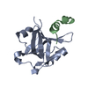 7xuvC  7xuwC  7xv0C  7xv1C 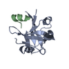 7xv4SC 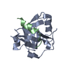 8jzyC 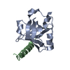 8k00C S: Starting model for refinement C: citing same article ( |
|---|---|
| Similar structure data |
-Links
 PDBj
PDBj


- Assembly
| Deposited unit | 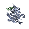
| ||||||||
|---|---|---|---|---|---|---|---|---|---|
| 1 |
| ||||||||
| Unit cell |
|
-Components
| #1: Protein | Mass: 13187.371 Da / Num. of mol.: 1 Source method: isolated from a genetically manipulated source Source: (gene. exp.) Homo sapiens (human) / Gene: RPA1, REPA1, RPA70 / Production host:  |
|---|---|
| #2: Protein/peptide | Mass: 2843.960 Da / Num. of mol.: 1 Source method: isolated from a genetically manipulated source Source: (gene. exp.) Homo sapiens (human) / Gene: ETAA1, ETAA16 / Production host: |
| #3: Water | ChemComp-HOH /  Mass: 18.015 Da / Num. of mol.: 139 / Source method: isolated from a natural source / Formula: H2O Mass: 18.015 Da / Num. of mol.: 139 / Source method: isolated from a natural source / Formula: H2O |
-Experimental details
-Experiment
| Experiment | Method: X-RAY DIFFRACTION / Number of used crystals: 1 |
|---|
- Sample preparation
| Crystal | Density Matthews: 2.34 Å3/Da / Density % sol: 47.48 % |
|---|---|
| Crystal grow | Temperature: 277 K / Method: vapor diffusion, hanging drop / Details: PEG 3350, Ammonium chloride |
-Data collection
| Diffraction | Mean temperature: 100 K / Serial crystal experiment: N |
|---|---|
| Diffraction source | Source: SYNCHROTRON / Site: SSRF / Beamline: BL19U1 / Wavelength: 0.9785 Å |
| Detector | Type: DECTRIS PILATUS 6M / Detector: PIXEL / Date: Aug 27, 2022 |
| Radiation | Protocol: SINGLE WAVELENGTH / Monochromatic (M) / Laue (L): M / Scattering type: x-ray |
| Radiation wavelength | Wavelength: 0.9785 Å / Relative weight: 1 |
| Reflection | Resolution: 1.5→39.5 Å / Num. obs: 42672 / % possible obs: 99.96 % / Redundancy: 9.4 % / CC1/2: 0.997 / Rmerge(I) obs: 0.05676 / Net I/σ(I): 26.24 |
| Reflection shell | Resolution: 1.5→1.54 Å / Rmerge(I) obs: 0.2213 / Num. unique obs: 2702 / CC1/2: 0.981 / Rpim(I) all: 0.07917 |
- Processing
| Software |
| ||||||||||||||||||||||||||||||||||||||||||||||||||||||||||||||||||||||||||||||||||||||||||||||||||||||||||||||||||||||||||||||||||||||||||||||||||||||||||||||||||||||||||||||||||||||||||||||||||||||||
|---|---|---|---|---|---|---|---|---|---|---|---|---|---|---|---|---|---|---|---|---|---|---|---|---|---|---|---|---|---|---|---|---|---|---|---|---|---|---|---|---|---|---|---|---|---|---|---|---|---|---|---|---|---|---|---|---|---|---|---|---|---|---|---|---|---|---|---|---|---|---|---|---|---|---|---|---|---|---|---|---|---|---|---|---|---|---|---|---|---|---|---|---|---|---|---|---|---|---|---|---|---|---|---|---|---|---|---|---|---|---|---|---|---|---|---|---|---|---|---|---|---|---|---|---|---|---|---|---|---|---|---|---|---|---|---|---|---|---|---|---|---|---|---|---|---|---|---|---|---|---|---|---|---|---|---|---|---|---|---|---|---|---|---|---|---|---|---|---|---|---|---|---|---|---|---|---|---|---|---|---|---|---|---|---|---|---|---|---|---|---|---|---|---|---|---|---|---|---|---|---|---|
| Refinement | Method to determine structure: MOLECULAR REPLACEMENT Starting model: 7XV4 Resolution: 1.5→39.5 Å / SU ML: 0.11 / Cross valid method: FREE R-VALUE / σ(F): 1.34 / Phase error: 22.6 / Stereochemistry target values: ML
| ||||||||||||||||||||||||||||||||||||||||||||||||||||||||||||||||||||||||||||||||||||||||||||||||||||||||||||||||||||||||||||||||||||||||||||||||||||||||||||||||||||||||||||||||||||||||||||||||||||||||
| Solvent computation | Shrinkage radii: 0.9 Å / VDW probe radii: 1.11 Å / Solvent model: FLAT BULK SOLVENT MODEL | ||||||||||||||||||||||||||||||||||||||||||||||||||||||||||||||||||||||||||||||||||||||||||||||||||||||||||||||||||||||||||||||||||||||||||||||||||||||||||||||||||||||||||||||||||||||||||||||||||||||||
| Refinement step | Cycle: LAST / Resolution: 1.5→39.5 Å
| ||||||||||||||||||||||||||||||||||||||||||||||||||||||||||||||||||||||||||||||||||||||||||||||||||||||||||||||||||||||||||||||||||||||||||||||||||||||||||||||||||||||||||||||||||||||||||||||||||||||||
| Refine LS restraints |
| ||||||||||||||||||||||||||||||||||||||||||||||||||||||||||||||||||||||||||||||||||||||||||||||||||||||||||||||||||||||||||||||||||||||||||||||||||||||||||||||||||||||||||||||||||||||||||||||||||||||||
| LS refinement shell |
| ||||||||||||||||||||||||||||||||||||||||||||||||||||||||||||||||||||||||||||||||||||||||||||||||||||||||||||||||||||||||||||||||||||||||||||||||||||||||||||||||||||||||||||||||||||||||||||||||||||||||
| Refinement TLS params. | Method: refined / Refine-ID: X-RAY DIFFRACTION
| ||||||||||||||||||||||||||||||||||||||||||||||||||||||||||||||||||||||||||||||||||||||||||||||||||||||||||||||||||||||||||||||||||||||||||||||||||||||||||||||||||||||||||||||||||||||||||||||||||||||||
| Refinement TLS group |
|
