 Movie
Movie Controller

[English] 日本語
 Yorodumi
- PDB-8jye: Crystal Structure of Intracellular B30.2 Domain of BTN3A1 and BTN... -
+ Open data

- Basic information
| Entry | Database: PDB / ID: 8jye | ||||||
|---|---|---|---|---|---|---|---|
| Title | Crystal Structure of Intracellular B30.2 Domain of BTN3A1 and BTN2A1 in Complex with HMBPP | ||||||
 Components Components | (Butyrophilin subfamily ...) x 2 | ||||||
 Keywords Keywords | SIGNALING PROTEIN / Butyrophilin | ||||||
| Function / homology |  Function and homology information Function and homology informationButyrophilin (BTN) family interactions / activated T cell proliferation / regulation of cytokine production / positive regulation of cytokine production / lipid metabolic process / positive regulation of type II interferon production / T cell receptor signaling pathway / adaptive immune response / signaling receptor binding / external side of plasma membrane / plasma membrane Similarity search - Function | ||||||
| Biological species |  Homo sapiens (human) Homo sapiens (human) | ||||||
| Method |  X-RAY DIFFRACTION / SYNCHROTRON / MOLECULAR REPLACEMENT / Resolution: 2.18 Å X-RAY DIFFRACTION / SYNCHROTRON / MOLECULAR REPLACEMENT / Resolution: 2.18 Å | ||||||
 Authors Authors | Yuan, L.J. / Yang, Y.Y. / Li, X. / Cai, N.N. / Chen, C.-C. / Guo, R.-T. / Zhang, Y.H. | ||||||
| Funding support |  China, 1items China, 1items
| ||||||
 Citation Citation | Journal: Nature / Year: 2023 Title: Phosphoantigens glue butyrophilin 3A1 and 2A1 to activate V gamma 9V delta 2 T cells. Authors: Yuan, L. / Ma, X. / Yang, Y. / Qu, Y. / Li, X. / Zhu, X. / Ma, W. / Duan, J. / Xue, J. / Yang, H. / Huang, J.W. / Yi, S. / Zhang, M. / Cai, N. / Zhang, L. / Ding, Q. / Lai, K. / Liu, C. / ...Authors: Yuan, L. / Ma, X. / Yang, Y. / Qu, Y. / Li, X. / Zhu, X. / Ma, W. / Duan, J. / Xue, J. / Yang, H. / Huang, J.W. / Yi, S. / Zhang, M. / Cai, N. / Zhang, L. / Ding, Q. / Lai, K. / Liu, C. / Zhang, L. / Liu, X. / Yao, Y. / Zhou, S. / Li, X. / Shen, P. / Chang, Q. / Malwal, S.R. / He, Y. / Li, W. / Chen, C. / Chen, C.C. / Oldfield, E. / Guo, R.T. / Zhang, Y. | ||||||
| History |
|
- Structure visualization
| Structure viewer | Molecule: MolmilJmol/JSmol |
|---|
- Downloads & links
-Download
| PDBx/mmCIF format | 8jye.cif.gz | 178.8 KB | Display | PDBx/mmCIF format |
|---|---|---|---|---|
| PDB format | pdb8jye.ent.gz | 138.6 KB | Display | PDB format |
| PDBx/mmJSON format | 8jye.json.gz | Tree view | PDBx/mmJSON format | |
| Others |  Other downloads Other downloads |
-Validation report
| Summary document | 8jye_validation.pdf.gz | 1 MB | Display | wwPDB validaton report |
|---|---|---|---|---|
| Full document | 8jye_full_validation.pdf.gz | 1.1 MB | Display | |
| Data in XML | 8jye_validation.xml.gz | 34.6 KB | Display | |
| Data in CIF | 8jye_validation.cif.gz | 48.4 KB | Display | |
| Arichive directory | https://data.pdbj.org/pub/pdb/validation_reports/jy/8jyeftp://data.pdbj.org/pub/pdb/validation_reports/jy/8jye | HTTPS FTP |
-Related structure data
| Related structure data |  8hjtC  8igtC 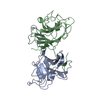 8ih4C 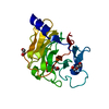 8ixvC 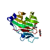 8izeC 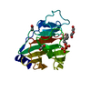 8izgC  8jy9C  8jyaC 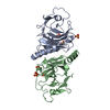 8jybC  8jycC 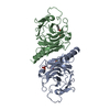 8jyfC  5zxkS S: Starting model for refinement C: citing same article ( |
|---|---|
| Similar structure data |
-Links
 PDBj
PDBj
- Assembly
| Deposited unit | 
| ||||||||
|---|---|---|---|---|---|---|---|---|---|
| 1 |
| ||||||||
| Unit cell |
|
-Components
-Butyrophilin subfamily ... , 2 types, 4 molecules ABCD
| #1: Protein | Mass: 24676.881 Da / Num. of mol.: 2 Source method: isolated from a genetically manipulated source Source: (gene. exp.) Homo sapiens (human) / Gene: BTN2A1, BT2.1, BTF1 / Production host:  #2: Protein | Mass: 22661.773 Da / Num. of mol.: 2 Source method: isolated from a genetically manipulated source Source: (gene. exp.) Homo sapiens (human) / Gene: BTN3A1 / Production host: |
|---|
-Non-polymers , 6 types, 406 molecules 


| #3: Chemical | ChemComp-PEG /  Mass: 106.120 Da / Num. of mol.: 4 / Source method: obtained synthetically / Formula: C4H10O3 Mass: 106.120 Da / Num. of mol.: 4 / Source method: obtained synthetically / Formula: C4H10O3#4: Chemical | ChemComp-EDO /  Mass: 62.068 Da / Num. of mol.: 4 / Source method: obtained synthetically / Formula: C2H6O2 Mass: 62.068 Da / Num. of mol.: 4 / Source method: obtained synthetically / Formula: C2H6O2#5: Chemical | ChemComp-1PE / |  Mass: 238.278 Da / Num. of mol.: 1 / Source method: obtained synthetically / Formula: C10H22O6 / Comment: precipitant*YM Mass: 238.278 Da / Num. of mol.: 1 / Source method: obtained synthetically / Formula: C10H22O6 / Comment: precipitant*YM#6: Chemical | ChemComp-PGE / |  Mass: 150.173 Da / Num. of mol.: 1 / Source method: obtained synthetically / Formula: C6H14O4 Mass: 150.173 Da / Num. of mol.: 1 / Source method: obtained synthetically / Formula: C6H14O4#7: Chemical |  Mass: 262.092 Da / Num. of mol.: 2 / Source method: obtained synthetically / Formula: C5H12O8P2 / Feature type: SUBJECT OF INVESTIGATION Mass: 262.092 Da / Num. of mol.: 2 / Source method: obtained synthetically / Formula: C5H12O8P2 / Feature type: SUBJECT OF INVESTIGATION#8: Water | ChemComp-HOH / | Mass: 18.015 Da / Num. of mol.: 394 / Source method: isolated from a natural source / Formula: H2O |
|---|
-Details
| Has ligand of interest | Y |
|---|
-Experimental details
-Experiment
| Experiment | Method: X-RAY DIFFRACTION / Number of used crystals: 1 |
|---|
- Sample preparation
| Crystal | Density Matthews: 3.61 Å3/Da / Density % sol: 65.91 % |
|---|---|
| Crystal grow | Temperature: 289 K / Method: vapor diffusion, sitting drop / Details: 42% PEG200, 0.1 M HEPES pH7.5 |
-Data collection
| Diffraction | Mean temperature: 100 K / Serial crystal experiment: N |
|---|---|
| Diffraction source | Source: SYNCHROTRON / Site: SSRF / Beamline: BL19U1 / Wavelength: 0.97883 Å |
| Detector | Type: DECTRIS PILATUS 6M / Detector: PIXEL / Date: Oct 22, 2020 |
| Radiation | Protocol: SINGLE WAVELENGTH / Monochromatic (M) / Laue (L): M / Scattering type: x-ray |
| Radiation wavelength | Wavelength: 0.97883 Å / Relative weight: 1 |
| Reflection | Resolution: 2.18→20.03 Å / Num. obs: 69590 / % possible obs: 99.41 % / Redundancy: 13.4 % / CC1/2: 0.999 / Rmerge(I) obs: 0.068 / Net I/σ(I): 21.9 |
| Reflection shell | Resolution: 2.18→2.25 Å / Num. unique obs: 6594 / CC1/2: 0.959 |
- Processing
| Software |
| ||||||||||||||||||||||||||||||||||||||||||||||||||||||||||||
|---|---|---|---|---|---|---|---|---|---|---|---|---|---|---|---|---|---|---|---|---|---|---|---|---|---|---|---|---|---|---|---|---|---|---|---|---|---|---|---|---|---|---|---|---|---|---|---|---|---|---|---|---|---|---|---|---|---|---|---|---|---|
| Refinement | Method to determine structure: MOLECULAR REPLACEMENT Starting model: 5ZXK Resolution: 2.18→20.03 Å / Cor.coef. Fo:Fc: 0.964 / Cor.coef. Fo:Fc free: 0.948 / SU B: 3.857 / SU ML: 0.1 / Cross valid method: THROUGHOUT / σ(F): 0 / ESU R: 0.161 / ESU R Free: 0.149 / Stereochemistry target values: MAXIMUM LIKELIHOOD Details: HYDROGENS HAVE BEEN ADDED IN THE RIDING POSITIONS U VALUES : REFINED INDIVIDUALLY
| ||||||||||||||||||||||||||||||||||||||||||||||||||||||||||||
| Solvent computation | Ion probe radii: 0.8 Å / Shrinkage radii: 0.8 Å / VDW probe radii: 1.2 Å / Solvent model: MASK | ||||||||||||||||||||||||||||||||||||||||||||||||||||||||||||
| Displacement parameters | Biso max: 120.05 Å2 / Biso mean: 44.08 Å2 / Biso min: 20.87 Å2
| ||||||||||||||||||||||||||||||||||||||||||||||||||||||||||||
| Refinement step | Cycle: final / Resolution: 2.18→20.03 Å
| ||||||||||||||||||||||||||||||||||||||||||||||||||||||||||||
| Refine LS restraints |
| ||||||||||||||||||||||||||||||||||||||||||||||||||||||||||||
| LS refinement shell | Resolution: 2.18→2.233 Å / Rfactor Rfree error: 0
|
