 Movie
Movie Controller

[English] 日本語
 Yorodumi
- PDB-8jx2: alpha-Hemolysin(G122S/K147R)-SpyTag/SpyCatcher head to head 14-mer -
+ Open data

- Basic information
| Entry | Database: PDB / ID: 8jx2 | ||||||||||||||||||||||||
|---|---|---|---|---|---|---|---|---|---|---|---|---|---|---|---|---|---|---|---|---|---|---|---|---|---|
| Title | alpha-Hemolysin(G122S/K147R)-SpyTag/SpyCatcher head to head 14-mer | ||||||||||||||||||||||||
 Components Components |
| ||||||||||||||||||||||||
 Keywords Keywords | TOXIN / pore / chimera protein / spy-catcher / spy-tag / dimer | ||||||||||||||||||||||||
| Function / homology |  Function and homology information Function and homology informationcytolysis in another organism / The NLRP3 inflammasome / Purinergic signaling in leishmaniasis infection / toxin activity / extracellular region / identical protein binding Similarity search - Function | ||||||||||||||||||||||||
| Biological species |   Staphylococcus aureus (bacteria) Staphylococcus aureus (bacteria) | ||||||||||||||||||||||||
| Method | ELECTRON MICROSCOPY / single particle reconstruction / cryo EM / Resolution: 2.2 Å | ||||||||||||||||||||||||
 Authors Authors | Ishii, Y. / Naito, K. / Yokoyama, T. / Tanaka, Y. / Matsuura, T. | ||||||||||||||||||||||||
| Funding support |  Japan, 1items Japan, 1items
| ||||||||||||||||||||||||
 Citation Citation | Journal: To Be Published Title: alpha-Hemolysin(G122S/K147R)-SpyTag/SpyCatcher head to head 14-mer Authors: Ishii, Y. / Naito, K. / Yokoyama, T. / Tanaka, Y. / Matsuura, T. | ||||||||||||||||||||||||
| History |
|
- Structure visualization
| Structure viewer | Molecule: MolmilJmol/JSmol |
|---|
- Downloads & links
-Download
| PDBx/mmCIF format | 8jx2.cif.gz | 717.6 KB | Display | PDBx/mmCIF format |
|---|---|---|---|---|
| PDB format | pdb8jx2.ent.gz | 596.5 KB | Display | PDB format |
| PDBx/mmJSON format | 8jx2.json.gz | Tree view | PDBx/mmJSON format | |
| Others |  Other downloads Other downloads |
-Validation report
| Arichive directory | https://data.pdbj.org/pub/pdb/validation_reports/jx/8jx2ftp://data.pdbj.org/pub/pdb/validation_reports/jx/8jx2 | HTTPS FTP |
|---|
-Related structure data
| Related structure data | 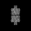 36688MC  8jx3C M: map data used to model this data C: citing same article ( |
|---|---|
| Similar structure data |

-Links
 PDBj
PDBj


- Assembly
| Deposited unit | 
|
|---|---|
| 1 |
|
-Components
| #1: Protein | Mass: 47003.504 Da / Num. of mol.: 7 / Mutation: G122S,K147R Source method: isolated from a genetically manipulated source Details: 300-412:SpyCatcher / Source: (gene. exp.) Staphylococcus aureus (bacteria) / Gene: hly, hla / Production host: #2: Protein | Mass: 36803.934 Da / Num. of mol.: 7 / Mutation: G122S,K147R Source method: isolated from a genetically manipulated source Details: 300-315:SpyTag / Source: (gene. exp.) Staphylococcus aureus (bacteria) / Gene: hly, hla / Production host: Has protein modification | N | |
|---|
-Experimental details
-Experiment
| Experiment | Method: ELECTRON MICROSCOPY |
|---|---|
| EM experiment | Aggregation state: PARTICLE / 3D reconstruction method: single particle reconstruction |
- Sample preparation
| Component | Name: heptameric alpha hemolysin pore dimerized by spy-catcher and spy-tag Type: COMPLEX / Entity ID: all / Source: RECOMBINANT | ||||||||||||
|---|---|---|---|---|---|---|---|---|---|---|---|---|---|
| Molecular weight | Experimental value: NO | ||||||||||||
| Source (natural) | Organism: Staphylococcus aureus (bacteria) | ||||||||||||
| Source (recombinant) | Organism: | ||||||||||||
| Buffer solution | pH: 7.6 / Details: 20mM HEPES-KOH[pH7.6], 50mM Potassium glutamate | ||||||||||||
| Buffer component |
| ||||||||||||
| Specimen | Conc.: 0.58 mg/ml / Embedding applied: NO / Shadowing applied: NO / Staining applied: NO / Vitrification applied: YES | ||||||||||||
| Specimen support | Grid type: Quantifoil R1.2/1.3 | ||||||||||||
| Vitrification | Instrument: FEI VITROBOT MARK IV / Cryogen name: ETHANE / Humidity: 100 % / Chamber temperature: 277 K |
- Electron microscopy imaging
| Microscopy | Model: JEOL CRYO ARM 300 |
|---|---|
| Electron gun | Electron source:  FIELD EMISSION GUN / Accelerating voltage: 300 kV / Illumination mode: FLOOD BEAM FIELD EMISSION GUN / Accelerating voltage: 300 kV / Illumination mode: FLOOD BEAM |
| Electron lens | Mode: BRIGHT FIELD / Nominal defocus max: 2500 nm / Nominal defocus min: 1500 nm |
| Image recording | Electron dose: 40 e/Å2 / Film or detector model: GATAN K3 (6k x 4k) |
- Processing
| EM software |
| ||||||||||||||||||||||||
|---|---|---|---|---|---|---|---|---|---|---|---|---|---|---|---|---|---|---|---|---|---|---|---|---|---|
| CTF correction | Type: PHASE FLIPPING AND AMPLITUDE CORRECTION | ||||||||||||||||||||||||
| 3D reconstruction | Resolution: 2.2 Å / Resolution method: FSC 0.143 CUT-OFF / Num. of particles: 349752 / Algorithm: FOURIER SPACE / Symmetry type: POINT | ||||||||||||||||||||||||
| Refine LS restraints |
|
