 Movie
Movie Controller

[English] 日本語
 Yorodumi
- EMDB-36689: alpha-Hemolysin(G122S/K147R/K237C)-SpyTag/SpyCatcher head to head... -
+ Open data

- Basic information
| Entry |  | |||||||||
|---|---|---|---|---|---|---|---|---|---|---|
| Title | alpha-Hemolysin(G122S/K147R/K237C)-SpyTag/SpyCatcher head to head 14-mer | |||||||||
 Map data Map data | B-factor sharpened map with B-factor value of -60. | |||||||||
 Sample Sample |
| |||||||||
 Keywords Keywords | pore / chimera protein / spy-catcher / spy-tag / dimer / TOXIN | |||||||||
| Function / homology |  Function and homology information Function and homology informationcytolysis in another organism / The NLRP3 inflammasome / Purinergic signaling in leishmaniasis infection / toxin activity / extracellular region / identical protein binding Similarity search - Function | |||||||||
| Biological species |   Staphylococcus aureus (bacteria) Staphylococcus aureus (bacteria) | |||||||||
| Method | single particle reconstruction / cryo EM / Resolution: 2.2 Å | |||||||||
 Authors Authors | Ishii Y / Naito K / Yokoyama T / Tanaka Y / Matsuura T | |||||||||
| Funding support |  Japan, 1 items Japan, 1 items
| |||||||||
 Citation Citation | Journal: To Be Published Title: alpha-Hemolysin(G122S/K147R)-SpyTag/SpyCatcher head to head 14-mer Authors: Ishii Y / Naito K / Yokoyama T / Tanaka Y / Matsuura T | |||||||||
| History |
|
- Structure visualization
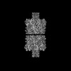
| Supplemental images |
|---|

- Downloads & links
-EMDB archive
| Map data | emd_36689.map.gz | 126.5 MB | EMDB map data format | |
|---|---|---|---|---|
| Header (meta data) | emd-36689-v30.xmlemd-36689.xml | 16.1 KB 16.1 KB | Display Display | EMDB header |
| FSC (resolution estimation) | emd_36689_fsc.xml | 14.9 KB | Display | FSC data file |
| Images |  emd_36689.png emd_36689.png | 82.6 KB | ||
| Filedesc metadata | emd-36689.cif.gz | 5.7 KB | ||
| Others | emd_36689_half_map_1.map.gzemd_36689_half_map_2.map.gz | 226.6 MB 226.6 MB | ||
| Archive directory |  http://ftp.pdbj.org/pub/emdb/structures/EMD-36689ftp://ftp.pdbj.org/pub/emdb/structures/EMD-36689 http://ftp.pdbj.org/pub/emdb/structures/EMD-36689ftp://ftp.pdbj.org/pub/emdb/structures/EMD-36689 | HTTPS FTP |
-Related structure data
| Related structure data |  8jx3MC 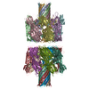 8jx2C M: atomic model generated by this map C: citing same article ( |
|---|---|
| Similar structure data |
-Links
| EMDB pages | EMDB (EBI/PDBe) / EMDataResource |
|---|---|
| Related items in Molecule of the Month |


-Map
| File | Download / File: emd_36689.map.gz / Format: CCP4 / Size: 244.1 MB / Type: IMAGE STORED AS FLOATING POINT NUMBER (4 BYTES) | ||||||||||||||||||||||||||||||||||||
|---|---|---|---|---|---|---|---|---|---|---|---|---|---|---|---|---|---|---|---|---|---|---|---|---|---|---|---|---|---|---|---|---|---|---|---|---|---|
| Annotation | B-factor sharpened map with B-factor value of -60. | ||||||||||||||||||||||||||||||||||||
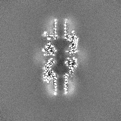
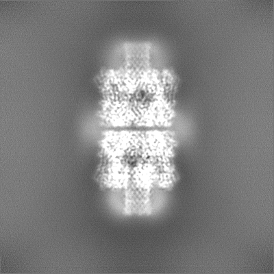
| Projections & slices | Image control
Images are generated by Spider. | ||||||||||||||||||||||||||||||||||||
| Voxel size | X=Y=Z: 0.788 Å | ||||||||||||||||||||||||||||||||||||
| Density |
| ||||||||||||||||||||||||||||||||||||
| Symmetry | Space group: 1 | ||||||||||||||||||||||||||||||||||||
| Details | EMDB XML:
|
 Z (Sec.)
 Y (Row.)
 X (Col.)


-Supplemental data
-Half map: One of the half maps.
| File | emd_36689_half_map_1.map | ||||||||||||
|---|---|---|---|---|---|---|---|---|---|---|---|---|---|
| Annotation | One of the half maps. | ||||||||||||
| Projections & Slices |
| ||||||||||||
| Density Histograms |


-Half map: One of the half maps.
| File | emd_36689_half_map_2.map | ||||||||||||
|---|---|---|---|---|---|---|---|---|---|---|---|---|---|
| Annotation | One of the half maps. | ||||||||||||
| Projections & Slices |
| ||||||||||||
| Density Histograms |


- Sample components
-Entire : heptameric alpha hemolysin (K237C) pore dimerized by spy-catcher ...
| Entire | Name: heptameric alpha hemolysin (K237C) pore dimerized by spy-catcher and spy-tag |
|---|---|
| Components |
|
-Supramolecule #1: heptameric alpha hemolysin (K237C) pore dimerized by spy-catcher ...
| Supramolecule | Name: heptameric alpha hemolysin (K237C) pore dimerized by spy-catcher and spy-tag type: complex / ID: 1 / Parent: 0 / Macromolecule list: all |
|---|---|
| Source (natural) | Organism: Staphylococcus aureus (bacteria) |
-Macromolecule #1: alpha hemolysin fused with spy-catcher
| Macromolecule | Name: alpha hemolysin fused with spy-catcher / type: protein_or_peptide / ID: 1 / Details: 300-412:SpyCatcher / Number of copies: 7 / Enantiomer: LEVO |
|---|---|
| Source (natural) | Organism: Staphylococcus aureus (bacteria) |
| Molecular weight | Theoretical: 46.977469 KDa |
| Recombinant expression | Organism: |
| Sequence | String: MADSDINIKT GTTDIGSNTT VKTGDLVTYD KENGMHKKVF YSFIDDKNHN KKLLVIRTKG TIAGQYRVYS EEGANKSGLA WPSAFKVQL QLPDNEVAQI SDYYPRNSID TKEYMSTLTY GFNSNVTGDD TGKIGGLIGA NVSIGHTLRY VQPDFKTILE S PTDKKVGW ...String: MADSDINIKT GTTDIGSNTT VKTGDLVTYD KENGMHKKVF YSFIDDKNHN KKLLVIRTKG TIAGQYRVYS EEGANKSGLA WPSAFKVQL QLPDNEVAQI SDYYPRNSID TKEYMSTLTY GFNSNVTGDD TGKIGGLIGA NVSIGHTLRY VQPDFKTILE S PTDKKVGW KVIFNNMVNQ NWGPYDRDSW NPVYGNQLFM KTRNGSMKAA DNFLDPNKAS SLLSSGFSPD FATVITMDRC AS KQQTNID VIYERVRDDY QLHWTSTNWK GTNTKDKWTD RSSERYKIDW EKEEMTNGSS GSVTTLSGLS GEQGPSGDMT TEE DSATHI KFSKRDEDGR ELAGATMELR DSSGKTISTW ISDGHVKDFY LYPGKYTFVE TAAPDGYEVA TPIEFTVNED GQVT VDGEA TEGDAHTGGS HHHHHH UniProtKB: Alpha-hemolysin |
-Macromolecule #2: alpha hemolysin fused with spy-tag
| Macromolecule | Name: alpha hemolysin fused with spy-tag / type: protein_or_peptide / ID: 2 / Details: 300-315:SpyTag / Number of copies: 7 / Enantiomer: LEVO |
|---|---|
| Source (natural) | Organism: Staphylococcus aureus (bacteria) |
| Molecular weight | Theoretical: 36.777898 KDa |
| Recombinant expression | Organism: |
| Sequence | String: MADSDINIKT GTTDIGSNTT VKTGDLVTYD KENGMHKKVF YSFIDDKNHN KKLLVIRTKG TIAGQYRVYS EEGANKSGLA WPSAFKVQL QLPDNEVAQI SDYYPRNSID TKEYMSTLTY GFNSNVTGDD TGKIGGLIGA NVSIGHTLRY VQPDFKTILE S PTDKKVGW ...String: MADSDINIKT GTTDIGSNTT VKTGDLVTYD KENGMHKKVF YSFIDDKNHN KKLLVIRTKG TIAGQYRVYS EEGANKSGLA WPSAFKVQL QLPDNEVAQI SDYYPRNSID TKEYMSTLTY GFNSNVTGDD TGKIGGLIGA NVSIGHTLRY VQPDFKTILE S PTDKKVGW KVIFNNMVNQ NWGPYDRDSW NPVYGNQLFM KTRNGSMKAA DNFLDPNKAS SLLSSGFSPD FATVITMDRC AS KQQTNID VIYERVRDDY QLHWTSTNWK GTNTKDKWTD RSSERYKIDW EKEEMTNGSS GSRGVPHIVM VDAYKRYKGG SHH HHHH UniProtKB: Alpha-hemolysin |
-Experimental details
-Structure determination
| Method | cryo EM |
|---|---|
 Processing Processing | single particle reconstruction |
| Aggregation state | particle |
-Sample preparation
| Concentration | 0.58 mg/mL | ||||||
|---|---|---|---|---|---|---|---|
| Buffer | pH: 7.6 Component:
Details: 20mM HEPES-KOH[pH7.6], 50mM Potassium glutamate | ||||||
| Grid | Model: Quantifoil R1.2/1.3 / Support film - Material: CARBON / Support film - topology: CONTINUOUS / Pretreatment - Type: GLOW DISCHARGE | ||||||
| Vitrification | Cryogen name: ETHANE / Chamber humidity: 100 % / Chamber temperature: 277 K / Instrument: FEI VITROBOT MARK IV |
- Electron microscopy
| Microscope | JEOL CRYO ARM 300 |
|---|---|
| Image recording | Film or detector model: GATAN K3 (6k x 4k) / Average electron dose: 40.0 e/Å2 |
| Electron beam | Acceleration voltage: 300 kV / Electron source:  FIELD EMISSION GUN FIELD EMISSION GUN |
| Electron optics | Illumination mode: FLOOD BEAM / Imaging mode: BRIGHT FIELD / Nominal defocus max: 2.4 µm / Nominal defocus min: 1.5 µm |

