National Natural Science Foundation of China (NSFC)
12034006
中国
National Science Foundation (NSF, China)
32071209
中国
National Natural Science Foundation of China (NSFC)
31971122
中国
National Natural Science Foundation of China (NSFC)
32200994
中国
引用
ジャーナル: J Mol Biol / 年: 2023 タイトル: Asymmetric Structure of Podophage GP4 Reveals a Novel Architecture of Three Types of Tail Fibers. 著者: Jing Zheng / Wenyuan Chen / Hao Xiao / Fan Yang / Jingdong Song / Lingpeng Cheng / Hongrong Liu / 要旨: Bacteriophage tail fibers (or called tail spikes) play a critical role in the early stage of infection by binding to the bacterial surface. Podophages with known structures usually possess one or two ...Bacteriophage tail fibers (or called tail spikes) play a critical role in the early stage of infection by binding to the bacterial surface. Podophages with known structures usually possess one or two types of fibers. Here, we resolved an asymmetric structure of the podophage GP4 to near-atomic resolution by cryo-EM. Our structure revealed a symmetry-mismatch relationship between the components of the GP4 tail with previously unseen topologies. In detail, two dodecameric adaptors (adaptors I and II), a hexameric nozzle, and a tail needle form a conserved tail body connected to a dodecameric portal occupying a unique vertex of the icosahedral head. However, five chain-like extended fibers (fiber I) and five tulip-like short fibers (fiber II) are anchored to a 15-fold symmetric fiber-tail adaptor, encircling the adaptor I, and six bamboo-like trimeric fibers (fiber III) are connected to the nozzle. Five fibers I, each composed of five dimers of the protein gp80 linked by an elongated rope protein, are attached to the five edges of the tail vertex of the icosahedral head. In this study, we identified a new structure of the podophage with three types of tail fibers, and such phages with different types of fibers may have a broad host range and/or infect host cells with considerably high efficiency, providing evolutionary advantages in harsh environments.
0: Portal protein 1: Putative tail fiber protein 2: Virion associated protein 3: Portal protein 4: Putative tail fiber protein 5: Virion associated protein 6: Portal protein 7: Putative tail fiber protein A: Virion-associated phage protein B: Virion-associated phage protein C: Virion-associated phage protein D: Virion-associated phage protein E: Virion-associated phage protein F: Virion-associated phage protein G: gp81 of phage GP4 H: gp81 of phage GP4 I: gp81 of phage GP4 J: gp81 of phage GP4 K: gp81 of phage GP4 L: gp81 of phage GP4 M: Putative tail fiber protein N: Putative tail fiber protein O: Putative tail fiber protein P: Putative tail fiber protein Q: Putative tail fiber protein R: Putative tail fiber protein S: gp81 of phage GP4 T: Virion associated protein U: Portal protein V: Putative tail fiber protein W: gp81 of phage GP4 X: Virion associated protein Y: Portal protein Z: Putative tail fiber protein a: gp81 of phage GP4 b: Virion associated protein c: Portal protein d: Putative tail fiber protein e: gp81 of phage GP4 f: Virion associated protein g: Portal protein h: Putative tail fiber protein i: gp81 of phage GP4 j: Virion associated protein k: Portal protein l: Putative tail fiber protein m: gp81 of phage GP4 n: Virion associated protein o: Portal protein p: Putative tail fiber protein q: Virion associated protein r: Portal protein s: Putative tail fiber protein t: Virion associated protein u: Portal protein v: Putative tail fiber protein w: Virion associated protein x: Portal protein y: Putative tail fiber protein z: Virion associated protein
 ムービー
ムービー コントローラー


 データを開く
 基本情報
基本情報 要素
要素 キーワード
キーワード 機能・相同性情報
 Ralstonia phage GP4 (ファージ)
 データ登録者
 中国, 4件
  引用
引用 構造の表示
構造の表示 ダウンロードとリンク
 その他のダウンロード


 PDBj
PDBj 集合体
集合体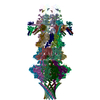
 試料調製
試料調製 電子顕微鏡撮影
 FIELD EMISSION GUN / 加速電圧: 200 kV / 照射モード: FLOOD BEAM
 解析
解析