 Movie
Movie Controller

[English] 日本語
 Yorodumi
- PDB-8jg5: Cryo-EM structure of the GI.4 Chiba VLP complexed with the CV-1A1... -
+ Open data

- Basic information
| Entry | Database: PDB / ID: 8jg5 | |||||||||||||||||||||||||||||||||||||||||||||||||||||||||||||||||||||||||||||||||||||||||||||
|---|---|---|---|---|---|---|---|---|---|---|---|---|---|---|---|---|---|---|---|---|---|---|---|---|---|---|---|---|---|---|---|---|---|---|---|---|---|---|---|---|---|---|---|---|---|---|---|---|---|---|---|---|---|---|---|---|---|---|---|---|---|---|---|---|---|---|---|---|---|---|---|---|---|---|---|---|---|---|---|---|---|---|---|---|---|---|---|---|---|---|---|---|---|---|
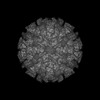
| Title | Cryo-EM structure of the GI.4 Chiba VLP complexed with the CV-1A1 Fv-clasp | |||||||||||||||||||||||||||||||||||||||||||||||||||||||||||||||||||||||||||||||||||||||||||||
 Components Components |
| |||||||||||||||||||||||||||||||||||||||||||||||||||||||||||||||||||||||||||||||||||||||||||||
 Keywords Keywords | VIRUS / NOROVIRUS / GI.4 / VIRUS LIKE PARTICLE / human monoclonal antibody / Fv-clasp | |||||||||||||||||||||||||||||||||||||||||||||||||||||||||||||||||||||||||||||||||||||||||||||
| Biological species |  Chiba virus Chiba virus Homo sapiens (human) Homo sapiens (human) | |||||||||||||||||||||||||||||||||||||||||||||||||||||||||||||||||||||||||||||||||||||||||||||
| Method | ELECTRON MICROSCOPY / single particle reconstruction / cryo EM / Resolution: 3.04 Å | |||||||||||||||||||||||||||||||||||||||||||||||||||||||||||||||||||||||||||||||||||||||||||||
 Authors Authors | Hosaka, T. / Katsura, K. / Kimura-Someya, T. / Someya, Y. / Shirouzu, M. | |||||||||||||||||||||||||||||||||||||||||||||||||||||||||||||||||||||||||||||||||||||||||||||
| Funding support |  Japan, 2items Japan, 2items
| |||||||||||||||||||||||||||||||||||||||||||||||||||||||||||||||||||||||||||||||||||||||||||||
 Citation Citation | Journal: J Virol / Year: 2024 Title: Structural analyses of the GI.4 norovirus by cryo-electron microscopy and X-ray crystallography revealing binding sites for human monoclonal antibodies. Authors: Tomomi Kimura-Someya / Kazushige Katsura / Miyuki Kato-Murayama / Toshiaki Hosaka / Tomomi Uchikubo-Kamo / Kentaro Ihara / Kazuharu Hanada / Shin Sato / Kazutaka Murayama / Michiyo Kataoka / ...Authors: Tomomi Kimura-Someya / Kazushige Katsura / Miyuki Kato-Murayama / Toshiaki Hosaka / Tomomi Uchikubo-Kamo / Kentaro Ihara / Kazuharu Hanada / Shin Sato / Kazutaka Murayama / Michiyo Kataoka / Mikako Shirouzu / Yuichi Someya / Abstract: Noroviruses are major causative agents of acute nonbacterial gastroenteritis in humans. There are neither antiviral therapeutic agents nor vaccines for noroviruses at this time. To evaluate the ...Noroviruses are major causative agents of acute nonbacterial gastroenteritis in humans. There are neither antiviral therapeutic agents nor vaccines for noroviruses at this time. To evaluate the potential usefulness of two previously isolated human monoclonal antibody fragments, CV-1A1 and CV-2F5, we first conducted a single-particle analysis to determine the cryo-electron microscopy structure of virus-like particles (VLPs) from the genogroup I genotype 4 (GI.4) Chiba strain uniformly coated with CV-1A1 fragments. The results revealed that the GI.4-specific CV-1A1 antibody bound to the P2 subdomain, in which amino acids are less conserved and variable. Interestingly, a part of the CV-1A1 intrudes into the histo-blood group antigen-binding site, suggesting that this antibody might exert neutralizing activity. Next, we determined the crystal structure of the protruding (P) domain of the capsid protein in the complex form with the CV-2F5 antibody fragment. Consistent with the cross-reactivity, the CV-2F5 bound to the P1 subdomain, which is rich in amino acids conserved among the GI strains, and moreover induced a disruption of Chiba VLPs. These results suggest that the broadly reactive CV-2F5 antibody can be used as both a universal detection reagent and an antiviral drug for GI noroviruses. IMPORTANCE: We conducted the structural analyses of the VP1 protein from the GI.4 Chiba norovirus to identify the binding sites of the previously isolated human monoclonal antibodies CV-1A1 and CV- ...IMPORTANCE: We conducted the structural analyses of the VP1 protein from the GI.4 Chiba norovirus to identify the binding sites of the previously isolated human monoclonal antibodies CV-1A1 and CV-2F5. The cryo-electron microscopy of the Chiba virus-like particles (VLPs) complexed with the Fv-clasp forms of GI.4-specific CV-1A1 revealed that this antibody binds to the highly variable P2 subdomain, suggesting that this antibody may have neutralizing ability against the GI.4 strains. X-ray crystallography revealed that the CV-2F5 antibody bound to the P1 subdomain, which is rich in conserved amino acids. This result is consistent with the ability of the CV-2F5 antibody to react with a wide variety of GI norovirus strains. It is also found that the CV-2F5 antibody caused a disruption of VLPs. Our findings, together with previous reports on the structures of VP1 proteins and VLPs, are expected to open a path for the structure-based development of antivirals and vaccines against norovirus disease. | |||||||||||||||||||||||||||||||||||||||||||||||||||||||||||||||||||||||||||||||||||||||||||||
| History |
|
- Structure visualization
| Structure viewer | Molecule:  MolmilJmol/JSmol MolmilJmol/JSmol |
|---|
- Downloads & links
-Download
| PDBx/mmCIF format | 8jg5.cif.gz | 404.1 KB | Display | PDBx/mmCIF format |
|---|---|---|---|---|
| PDB format | pdb8jg5.ent.gz | 327.3 KB | Display | PDB format |
| PDBx/mmJSON format | 8jg5.json.gz | Tree view | PDBx/mmJSON format | |
| Others |  Other downloads Other downloads |
-Validation report
| Arichive directory | https://data.pdbj.org/pub/pdb/validation_reports/jg/8jg5ftp://data.pdbj.org/pub/pdb/validation_reports/jg/8jg5 | HTTPS FTP |
|---|
-Related structure data

-Links
 PDBj
PDBj

- Assembly
| Deposited unit | 
|
|---|---|
| 1 | x 60
|
| 2 |
|
| 3 | x 5
|
| 4 | x 6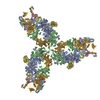
|
| 5 | 
|
| Symmetry | Point symmetry: (Schoenflies symbol: I (icosahedral)) |
-Components
| #1: Protein | Mass: 58108.332 Da / Num. of mol.: 3 Source method: isolated from a genetically manipulated source Details: GenBank ID AB042808 / Source: (gene. exp.) Chiba virus / Strain: GI/Human/Japan/Chiba 407/1987 / Gene: ORF2 / Production host:   Spodoptera frugiperda (fall armyworm) Spodoptera frugiperda (fall armyworm)#2: Antibody | Mass: 19847.254 Da / Num. of mol.: 2 Source method: isolated from a genetically manipulated source Details: VH-SARAH chimera of linker (residues (-6)-0), VH (residues 1-118), and SARAH (residues 119-171) Source: (gene. exp.) Homo sapiens (human) / Production host:  #3: Antibody | Mass: 18193.150 Da / Num. of mol.: 2 Source method: isolated from a genetically manipulated source Details: VL-SARAH chimera of linker (residues (-6)-0), VL (residues 1-112), and SARAH (residues 113-162) Source: (gene. exp.) Homo sapiens (human) / Production host: Has protein modification | Y | |
|---|
-Experimental details
-Experiment
| Experiment | Method: ELECTRON MICROSCOPY |
|---|---|
| EM experiment | Aggregation state: PARTICLE / 3D reconstruction method: single particle reconstruction |
- Sample preparation
| Component | Name: Viral protein 1 with its antibody fragments / Type: COMPLEX / Entity ID: all / Source: MULTIPLE SOURCES |
|---|---|
| Source (natural) | Organism: Chiba virus / Strain: GI/Human/Japan/Chiba 407/1987 |
| Source (recombinant) | Organism: Spodoptera frugiperda (fall armyworm) |
| Buffer solution | pH: 8 |
| Specimen | Embedding applied: NO / Shadowing applied: NO / Staining applied: NO / Vitrification applied: YES |
| Vitrification | Cryogen name: ETHANE |
- Electron microscopy imaging
| Experimental equipment |  Model: Talos Arctica / Image courtesy: FEI Company |
|---|---|
| Microscopy | Model: FEI TECNAI ARCTICA |
| Electron gun | Electron source:  FIELD EMISSION GUN / Accelerating voltage: 200 kV / Illumination mode: FLOOD BEAM FIELD EMISSION GUN / Accelerating voltage: 200 kV / Illumination mode: FLOOD BEAM |
| Electron lens | Mode: BRIGHT FIELD / Nominal defocus max: 1600 nm / Nominal defocus min: 800 nm |
| Image recording | Electron dose: 50 e/Å2 / Film or detector model: GATAN K2 SUMMIT (4k x 4k) |
- Processing
| Software | Name: PHENIX / Version: 1.20.1_4487: / Classification: refinement | ||||||||||||||||||||||||
|---|---|---|---|---|---|---|---|---|---|---|---|---|---|---|---|---|---|---|---|---|---|---|---|---|---|
| EM software | Name: PHENIX / Category: model refinement | ||||||||||||||||||||||||
| CTF correction | Type: PHASE FLIPPING AND AMPLITUDE CORRECTION | ||||||||||||||||||||||||
| Symmetry | Point symmetry: I (icosahedral) | ||||||||||||||||||||||||
| 3D reconstruction | Resolution: 3.04 Å / Resolution method: FSC 0.143 CUT-OFF / Num. of particles: 47125 / Symmetry type: POINT | ||||||||||||||||||||||||
| Refine LS restraints |
|
