Japan Agency for Medical Research and Development (AMED)
Japan
Citation
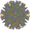
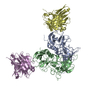
Journal: J Virol / Year: 2024 Title: Structural analyses of the GI.4 norovirus by cryo-electron microscopy and X-ray crystallography revealing binding sites for human monoclonal antibodies. Authors: Tomomi Kimura-Someya / Kazushige Katsura / Miyuki Kato-Murayama / Toshiaki Hosaka / Tomomi Uchikubo-Kamo / Kentaro Ihara / Kazuharu Hanada / Shin Sato / Kazutaka Murayama / Michiyo Kataoka / ...Authors: Tomomi Kimura-Someya / Kazushige Katsura / Miyuki Kato-Murayama / Toshiaki Hosaka / Tomomi Uchikubo-Kamo / Kentaro Ihara / Kazuharu Hanada / Shin Sato / Kazutaka Murayama / Michiyo Kataoka / Mikako Shirouzu / Yuichi Someya / Abstract: Noroviruses are major causative agents of acute nonbacterial gastroenteritis in humans. There are neither antiviral therapeutic agents nor vaccines for noroviruses at this time. To evaluate the ...Noroviruses are major causative agents of acute nonbacterial gastroenteritis in humans. There are neither antiviral therapeutic agents nor vaccines for noroviruses at this time. To evaluate the potential usefulness of two previously isolated human monoclonal antibody fragments, CV-1A1 and CV-2F5, we first conducted a single-particle analysis to determine the cryo-electron microscopy structure of virus-like particles (VLPs) from the genogroup I genotype 4 (GI.4) Chiba strain uniformly coated with CV-1A1 fragments. The results revealed that the GI.4-specific CV-1A1 antibody bound to the P2 subdomain, in which amino acids are less conserved and variable. Interestingly, a part of the CV-1A1 intrudes into the histo-blood group antigen-binding site, suggesting that this antibody might exert neutralizing activity. Next, we determined the crystal structure of the protruding (P) domain of the capsid protein in the complex form with the CV-2F5 antibody fragment. Consistent with the cross-reactivity, the CV-2F5 bound to the P1 subdomain, which is rich in amino acids conserved among the GI strains, and moreover induced a disruption of Chiba VLPs. These results suggest that the broadly reactive CV-2F5 antibody can be used as both a universal detection reagent and an antiviral drug for GI noroviruses. IMPORTANCE: We conducted the structural analyses of the VP1 protein from the GI.4 Chiba norovirus to identify the binding sites of the previously isolated human monoclonal antibodies CV-1A1 and CV- ...IMPORTANCE: We conducted the structural analyses of the VP1 protein from the GI.4 Chiba norovirus to identify the binding sites of the previously isolated human monoclonal antibodies CV-1A1 and CV-2F5. The cryo-electron microscopy of the Chiba virus-like particles (VLPs) complexed with the Fv-clasp forms of GI.4-specific CV-1A1 revealed that this antibody binds to the highly variable P2 subdomain, suggesting that this antibody may have neutralizing ability against the GI.4 strains. X-ray crystallography revealed that the CV-2F5 antibody bound to the P1 subdomain, which is rich in conserved amino acids. This result is consistent with the ability of the CV-2F5 antibody to react with a wide variety of GI norovirus strains. It is also found that the CV-2F5 antibody caused a disruption of VLPs. Our findings, together with previous reports on the structures of VP1 proteins and VLPs, are expected to open a path for the structure-based development of antivirals and vaccines against norovirus disease.
Mass: 33895.895 Da / Num. of mol.: 3 Source method: isolated from a genetically manipulated source Source: (gene. exp.) Chiba virus / Strain: GI/Human/Japan/Chiba 407/1987 / Production host: Escherichia coli (E. coli) / References: UniProt: Q9DU46
#2: Antibody
scFvfragment
Mass: 27334.945 Da / Num. of mol.: 2 Source method: isolated from a genetically manipulated source Source: (gene. exp.) Homo sapiens (human) / Production host: Escherichia coli (E. coli)
In the structure databanks used in Yorodumi, some data are registered as the other names, "COVID-19 virus" and "2019-nCoV". Here are the details of the virus and the list of structure data.
Jan 31, 2019. EMDB accession codes are about to change! (news from PDBe EMDB page)
EMDB accession codes are about to change! (news from PDBe EMDB page)
The allocation of 4 digits for EMDB accession codes will soon come to an end. Whilst these codes will remain in use, new EMDB accession codes will include an additional digit and will expand incrementally as the available range of codes is exhausted. The current 4-digit format prefixed with “EMD-” (i.e. EMD-XXXX) will advance to a 5-digit format (i.e. EMD-XXXXX), and so on. It is currently estimated that the 4-digit codes will be depleted around Spring 2019, at which point the 5-digit format will come into force.
The EM Navigator/Yorodumi systems omit the EMD- prefix.
Related info.:Q: What is EMD? / ID/Accession-code notation in Yorodumi/EM Navigator
Yorodumi is a browser for structure data from EMDB, PDB, SASBDB, etc.
This page is also the successor to EM Navigator detail page, and also detail information page/front-end page for Omokage search.
The word "yorodu" (or yorozu) is an old Japanese word meaning "ten thousand". "mi" (miru) is to see.
Related info.:EMDB / PDB / SASBDB / Comparison of 3 databanks / Yorodumi Search / Aug 31, 2016. New EM Navigator & Yorodumi / Yorodumi Papers / Jmol/JSmol / Function and homology information / Changes in new EM Navigator and Yorodumi
 Movie
Movie Controller

 Yorodumi
 Open data
 Basic information
 Components
 Keywords
 Function and homology information
 Chiba virus
 Homo sapiens (human)
 X-RAY DIFFRACTION /
  Authors
 Japan, 1items
  Citation
 Structure visualization
 Downloads & links
 Other downloads
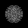
 PDBj
PDBj


 Assembly
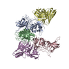


 Mass: 18.015 Da / Num. of mol.: 177 / Source method: isolated from a natural source / Formula: H2O
 Sample preparation
 Processing