 Movie
Movie Controller

[English] 日本語
 Yorodumi
- PDB-8je9: Crystal structure of CGL1 from Crassostrea gigas, mannobiose-boun... -
+ Open data

- Basic information
| Entry | Database: PDB / ID: 8je9 | ||||||
|---|---|---|---|---|---|---|---|
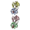
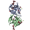
| Title | Crystal structure of CGL1 from Crassostrea gigas, mannobiose-bound form (CGL1/Man(alpha)1-2Man) | ||||||
 Components Components | Natterin-3 | ||||||
 Keywords Keywords | SUGAR BINDING PROTEIN / complex structure | ||||||
| Function / homology | DM9 repeat / DM9 repeat / Repeats found in Drosophila proteins. / metal ion binding / ACETIC ACID / CACODYLATE ION / alpha-D-mannopyranose / Natterin-3 Function and homology information Function and homology information | ||||||
| Biological species |  Crassostrea gigas (Pacific oyster) Crassostrea gigas (Pacific oyster) | ||||||
| Method |  X-RAY DIFFRACTION / SYNCHROTRON / MOLECULAR REPLACEMENT / Resolution: 1 Å X-RAY DIFFRACTION / SYNCHROTRON / MOLECULAR REPLACEMENT / Resolution: 1 Å | ||||||
 Authors Authors | Unno, H. / Hatakeyama, T. | ||||||
| Funding support |  Japan, 1items Japan, 1items
| ||||||
 Citation Citation | Journal: J.Biochem. / Year: 2023 Title: Mannose oligosaccharide recognition of CGL1, a mannose-specific lectin containing DM9 motifs from Crassostrea gigas, revealed by X-ray crystallographic analysis. Authors: Hatakeyama, T. / Masuda, K. / Kudo, M. / Tanaka, K. / Takeuchi, A. / Unno, H. | ||||||
| History |
|
- Structure visualization
| Structure viewer | Molecule: MolmilJmol/JSmol |
|---|
- Downloads & links
-Download
| PDBx/mmCIF format | 8je9.cif.gz | 283.4 KB | Display | PDBx/mmCIF format |
|---|---|---|---|---|
| PDB format | pdb8je9.ent.gz | 215.5 KB | Display | PDB format |
| PDBx/mmJSON format | 8je9.json.gz | Tree view | PDBx/mmJSON format | |
| Others |  Other downloads Other downloads |
-Validation report
| Arichive directory | https://data.pdbj.org/pub/pdb/validation_reports/je/8je9ftp://data.pdbj.org/pub/pdb/validation_reports/je/8je9 | HTTPS FTP |
|---|
-Related structure data
-Links
 PDBj
PDBj


- Assembly
| Deposited unit | 
| ||||||||
|---|---|---|---|---|---|---|---|---|---|
| 1 | 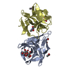
| ||||||||
| 2 | 
| ||||||||
| Unit cell |
|
-Components
-Protein , 1 types, 4 molecules ABCD
| #1: Protein | Mass: 15578.734 Da / Num. of mol.: 4 / Source method: isolated from a natural source / Source: (natural) Crassostrea gigas (Pacific oyster) / References: UniProt: K1QRB6 |
|---|
-Sugars , 2 types, 3 molecules 
| #2: Polysaccharide | Source method: isolated from a genetically manipulated source #4: Sugar | ChemComp-MAN / |  Type: D-saccharide, alpha linking / Mass: 180.156 Da / Num. of mol.: 1 / Source method: obtained synthetically / Formula: C6H12O6 / Feature type: SUBJECT OF INVESTIGATION Type: D-saccharide, alpha linking / Mass: 180.156 Da / Num. of mol.: 1 / Source method: obtained synthetically / Formula: C6H12O6 / Feature type: SUBJECT OF INVESTIGATION |
|---|
-Non-polymers , 3 types, 806 molecules 


| #3: Chemical |  Mass: 60.052 Da / Num. of mol.: 3 / Source method: obtained synthetically / Formula: C2H4O2 Mass: 60.052 Da / Num. of mol.: 3 / Source method: obtained synthetically / Formula: C2H4O2#5: Chemical | ChemComp-CAC / |  Mass: 136.989 Da / Num. of mol.: 1 / Source method: obtained synthetically / Formula: C2H6AsO2 Mass: 136.989 Da / Num. of mol.: 1 / Source method: obtained synthetically / Formula: C2H6AsO2#6: Water | ChemComp-HOH / | Mass: 18.015 Da / Num. of mol.: 802 / Source method: isolated from a natural source / Formula: H2O |
|---|
-Details
| Has ligand of interest | Y |
|---|
-Experimental details
-Experiment
| Experiment | Method: X-RAY DIFFRACTION / Number of used crystals: 1 |
|---|
- Sample preparation
| Crystal | Density Matthews: 2.03 Å3/Da / Density % sol: 39.27 % |
|---|---|
| Crystal grow | Temperature: 293 K / Method: vapor diffusion Details: 0.2 M magnesium acetate, 0.1 M sodium cacodylate (pH 6.5), 20% (w/v) PEG 8000 |
-Data collection
| Diffraction | Mean temperature: 90 K / Serial crystal experiment: N |
|---|---|
| Diffraction source | Source: SYNCHROTRON / Site: Photon Factory / Beamline: BL-5A / Wavelength: 1 Å |
| Detector | Type: DECTRIS PILATUS3 S 6M / Detector: PIXEL / Date: Dec 2, 2021 |
| Radiation | Protocol: SINGLE WAVELENGTH / Monochromatic (M) / Laue (L): M / Scattering type: x-ray |
| Radiation wavelength | Wavelength: 1 Å / Relative weight: 1 |
| Reflection | Resolution: 1→39.8 Å / Num. obs: 229413 / % possible obs: 85.9 % / Redundancy: 6.8 % / CC1/2: 0.999 / Rmerge(I) obs: 0.056 / Net I/σ(I): 18.6 |
| Reflection shell | Resolution: 1→1.02 Å / Num. unique obs: 4020 / CC1/2: 0.872 |
- Processing
| Software |
| |||||||||||||||||||||||||||||||||||||||||||||||||||||||||||||||||||||||||||||||||||||||||||||||||||||||||||||||||||||||||||||||||||||||||||||||||||||||||||||||||||||||||||||||||||||||||||||||||||||||||||||||||||||||||||||||||||||||
|---|---|---|---|---|---|---|---|---|---|---|---|---|---|---|---|---|---|---|---|---|---|---|---|---|---|---|---|---|---|---|---|---|---|---|---|---|---|---|---|---|---|---|---|---|---|---|---|---|---|---|---|---|---|---|---|---|---|---|---|---|---|---|---|---|---|---|---|---|---|---|---|---|---|---|---|---|---|---|---|---|---|---|---|---|---|---|---|---|---|---|---|---|---|---|---|---|---|---|---|---|---|---|---|---|---|---|---|---|---|---|---|---|---|---|---|---|---|---|---|---|---|---|---|---|---|---|---|---|---|---|---|---|---|---|---|---|---|---|---|---|---|---|---|---|---|---|---|---|---|---|---|---|---|---|---|---|---|---|---|---|---|---|---|---|---|---|---|---|---|---|---|---|---|---|---|---|---|---|---|---|---|---|---|---|---|---|---|---|---|---|---|---|---|---|---|---|---|---|---|---|---|---|---|---|---|---|---|---|---|---|---|---|---|---|---|---|---|---|---|---|---|---|---|---|---|---|---|---|---|---|---|---|
| Refinement | Method to determine structure: MOLECULAR REPLACEMENT / Resolution: 1→39.778 Å / Cor.coef. Fo:Fc: 0.975 / Cor.coef. Fo:Fc free: 0.971 / WRfactor Rfree: 0.206 / WRfactor Rwork: 0.192 / Average fsc free: 0.9723 / Average fsc work: 0.976 / Cross valid method: FREE R-VALUE / ESU R: 0.033 / ESU R Free: 0.031 Details: Hydrogens have been added in their riding positions
| |||||||||||||||||||||||||||||||||||||||||||||||||||||||||||||||||||||||||||||||||||||||||||||||||||||||||||||||||||||||||||||||||||||||||||||||||||||||||||||||||||||||||||||||||||||||||||||||||||||||||||||||||||||||||||||||||||||||
| Solvent computation | Ion probe radii: 0.8 Å / Shrinkage radii: 0.8 Å / VDW probe radii: 1.2 Å / Solvent model: MASK BULK SOLVENT | |||||||||||||||||||||||||||||||||||||||||||||||||||||||||||||||||||||||||||||||||||||||||||||||||||||||||||||||||||||||||||||||||||||||||||||||||||||||||||||||||||||||||||||||||||||||||||||||||||||||||||||||||||||||||||||||||||||||
| Displacement parameters | Biso mean: 10.872 Å2
| |||||||||||||||||||||||||||||||||||||||||||||||||||||||||||||||||||||||||||||||||||||||||||||||||||||||||||||||||||||||||||||||||||||||||||||||||||||||||||||||||||||||||||||||||||||||||||||||||||||||||||||||||||||||||||||||||||||||
| Refinement step | Cycle: LAST / Resolution: 1→39.778 Å
| |||||||||||||||||||||||||||||||||||||||||||||||||||||||||||||||||||||||||||||||||||||||||||||||||||||||||||||||||||||||||||||||||||||||||||||||||||||||||||||||||||||||||||||||||||||||||||||||||||||||||||||||||||||||||||||||||||||||
| Refine LS restraints |
| |||||||||||||||||||||||||||||||||||||||||||||||||||||||||||||||||||||||||||||||||||||||||||||||||||||||||||||||||||||||||||||||||||||||||||||||||||||||||||||||||||||||||||||||||||||||||||||||||||||||||||||||||||||||||||||||||||||||
| LS refinement shell | Refine-ID: X-RAY DIFFRACTION / Total num. of bins used: 20
|
