 Movie
Movie Controller

[English] 日本語
 Yorodumi
- PDB-8ja7: Cryo-EM structure of Mycobacterium tuberculosis LpqY-SugABC in co... -
+ Open data

- Basic information
| Entry | Database: PDB / ID: 8ja7 | |||||||||||||||||||||||||||||||||
|---|---|---|---|---|---|---|---|---|---|---|---|---|---|---|---|---|---|---|---|---|---|---|---|---|---|---|---|---|---|---|---|---|---|---|
| Title | Cryo-EM structure of Mycobacterium tuberculosis LpqY-SugABC in complex with trehalose | |||||||||||||||||||||||||||||||||
 Components Components |
| |||||||||||||||||||||||||||||||||
 Keywords Keywords | MEMBRANE PROTEIN / ABC transporter | |||||||||||||||||||||||||||||||||
| Function / homology |  Function and homology information Function and homology informationABC-type carbohydrate transporter activity / ATP-binding cassette (ABC) transporter complex, transmembrane substrate-binding subunit-containing / Translocases; Catalysing the translocation of carbohydrates and their derivatives; Linked to the hydrolysis of a nucleoside triphosphate / trehalose transmembrane transporter activity / trehalose transport / biological process involved in interaction with host / ATP-binding cassette (ABC) transporter complex, substrate-binding subunit-containing / ATP-binding cassette (ABC) transporter complex / transmembrane transport / periplasmic space ...ABC-type carbohydrate transporter activity / ATP-binding cassette (ABC) transporter complex, transmembrane substrate-binding subunit-containing / Translocases; Catalysing the translocation of carbohydrates and their derivatives; Linked to the hydrolysis of a nucleoside triphosphate / trehalose transmembrane transporter activity / trehalose transport / biological process involved in interaction with host / ATP-binding cassette (ABC) transporter complex, substrate-binding subunit-containing / ATP-binding cassette (ABC) transporter complex / transmembrane transport / periplasmic space / protein homodimerization activity / ATP hydrolysis activity / ATP binding / identical protein binding / plasma membrane Similarity search - Function | |||||||||||||||||||||||||||||||||
| Biological species |  Mycobacterium tuberculosis H37Rv (bacteria) Mycobacterium tuberculosis H37Rv (bacteria) | |||||||||||||||||||||||||||||||||
| Method | ELECTRON MICROSCOPY / single particle reconstruction / cryo EM / Resolution: 3.02 Å | |||||||||||||||||||||||||||||||||
 Authors Authors | Zhang, B. / Liang, J. / Rao, Z. | |||||||||||||||||||||||||||||||||
| Funding support |  China, 2items China, 2items
| |||||||||||||||||||||||||||||||||
 Citation Citation | Journal: Proc Natl Acad Sci U S A / Year: 2023 Title: Molecular recognition of trehalose and trehalose analogues by LpqY-SugABC. Authors: Jingxi Liang / Fengjiang Liu / Peng Xu / Wei Shangguan / Tianyu Hu / Shule Wang / Xiaolin Yang / Zhiqi Xiong / Xiuna Yang / Luke W Guddat / Biao Yu / Zihe Rao / Bing Zhang /  Abstract: Trehalose plays a crucial role in the survival and virulence of the deadly human pathogen (). The type I ATP-binding cassette (ABC) transporter LpqY-SugABC is the sole pathway for trehalose to enter ...Trehalose plays a crucial role in the survival and virulence of the deadly human pathogen (). The type I ATP-binding cassette (ABC) transporter LpqY-SugABC is the sole pathway for trehalose to enter . The substrate-binding protein, LpqY, which forms a stable complex with the translocator SugABC, recognizes and captures trehalose and its analogues in the periplasmic space, but the precise molecular mechanism for this process is still not well understood. This study reports a 3.02-Å cryoelectron microscopy structure of trehalose-bound LpqY-SugABC in the pretranslocation state, a crystal structure of LpqY in a closed form with trehalose bound and five crystal structures of LpqY in complex with different trehalose analogues. These structures, accompanied by substrate-stimulated ATPase activity data, reveal how LpqY recognizes and binds trehalose and its analogues, and highlight the flexibility in the substrate binding pocket of LpqY. These data provide critical insights into the design of trehalose analogues that could serve as potential molecular probe tools or as anti-TB drugs. | |||||||||||||||||||||||||||||||||
| History |
|
- Structure visualization
| Structure viewer | Molecule: MolmilJmol/JSmol |
|---|
- Downloads & links
-Download
| PDBx/mmCIF format | 8ja7.cif.gz | 307.2 KB | Display | PDBx/mmCIF format |
|---|---|---|---|---|
| PDB format | pdb8ja7.ent.gz | 247 KB | Display | PDB format |
| PDBx/mmJSON format | 8ja7.json.gz | Tree view | PDBx/mmJSON format | |
| Others |  Other downloads Other downloads |
-Validation report
| Arichive directory | https://data.pdbj.org/pub/pdb/validation_reports/ja/8ja7ftp://data.pdbj.org/pub/pdb/validation_reports/ja/8ja7 | HTTPS FTP |
|---|
-Related structure data
| Related structure data |  36125MC 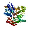 8ja8C  8ja9C  8jaaC 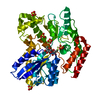 8jabC 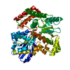 8jacC  8jadC M: map data used to model this data C: citing same article ( |
|---|---|
| Similar structure data |
-Links
 PDBj
PDBj


- Assembly
| Deposited unit | 
|
|---|---|
| 1 |
|
-Components
| #1: Protein | Mass: 33041.809 Da / Num. of mol.: 1 Source method: isolated from a genetically manipulated source Source: (gene. exp.) Mycobacterium tuberculosis H37Rv (bacteria)Gene: sugA Production host: Mycolicibacterium smegmatis MC2 155 (bacteria)References: UniProt: P9WG03 | ||||||
|---|---|---|---|---|---|---|---|
| #2: Protein | Mass: 29145.537 Da / Num. of mol.: 1 Source method: isolated from a genetically manipulated source Source: (gene. exp.) Mycobacterium tuberculosis H37Rv (bacteria)Gene: sugB Production host: Mycolicibacterium smegmatis MC2 155 (bacteria)References: UniProt: P9WG01 | ||||||
| #3: Protein | Mass: 49803.074 Da / Num. of mol.: 1 Source method: isolated from a genetically manipulated source Source: (gene. exp.) Mycobacterium tuberculosis H37Rv (bacteria)Gene: lpqY Production host: Mycolicibacterium smegmatis MC2 155 (bacteria)References: UniProt: P9WGU9 | ||||||
| #4: Protein | Mass: 42964.078 Da / Num. of mol.: 2 Source method: isolated from a genetically manipulated source Source: (gene. exp.) Mycobacterium tuberculosis H37Rv (bacteria)Gene: sugC Production host: Mycolicibacterium smegmatis MC2 155 (bacteria)References: UniProt: P9WQI3, Translocases; Catalysing the translocation of carbohydrates and their derivatives; Linked to the hydrolysis of a nucleoside triphosphate #5: Polysaccharide | alpha-D-glucopyranose-(1-1)-alpha-D-glucopyranose | Has ligand of interest | Y | Has protein modification | N | |
-Experimental details
-Experiment
| Experiment | Method: ELECTRON MICROSCOPY |
|---|---|
| EM experiment | Aggregation state: PARTICLE / 3D reconstruction method: single particle reconstruction |
- Sample preparation
| Component | Name: LpqY-SugABC / Type: COMPLEX / Entity ID: #1-#4 / Source: RECOMBINANT |
|---|---|
| Source (natural) | Organism: Mycobacterium tuberculosis H37Rv (bacteria) |
| Source (recombinant) | Organism: Mycolicibacterium smegmatis MC2 155 (bacteria) |
| Buffer solution | pH: 7.5 |
| Specimen | Embedding applied: NO / Shadowing applied: NO / Staining applied: NO / Vitrification applied: YES |
| Vitrification | Cryogen name: ETHANE |
- Electron microscopy imaging
| Microscopy | Model: FEI TITAN |
|---|---|
| Electron gun | Electron source:  FIELD EMISSION GUN / Accelerating voltage: 300 kV / Illumination mode: FLOOD BEAM FIELD EMISSION GUN / Accelerating voltage: 300 kV / Illumination mode: FLOOD BEAM |
| Electron lens | Mode: BRIGHT FIELD / Nominal defocus max: 2000 nm / Nominal defocus min: 1200 nm |
| Image recording | Electron dose: 60 e/Å2 / Film or detector model: GATAN K3 (6k x 4k) |
- Processing
| Software | Name: PHENIX / Version: 1.16_3549: / Classification: refinement | ||||||||||||||||||||||||
|---|---|---|---|---|---|---|---|---|---|---|---|---|---|---|---|---|---|---|---|---|---|---|---|---|---|
| EM software | Name: PHENIX / Category: model refinement | ||||||||||||||||||||||||
| CTF correction | Type: PHASE FLIPPING AND AMPLITUDE CORRECTION | ||||||||||||||||||||||||
| 3D reconstruction | Resolution: 3.02 Å / Resolution method: FSC 0.143 CUT-OFF / Num. of particles: 50775 / Symmetry type: POINT | ||||||||||||||||||||||||
| Refine LS restraints |
|
