 Movie
Movie Controller

[English] 日本語
 Yorodumi
- PDB-8j92: Cryo-EM structure of nucleosome containing Arabidopsis thaliana H2A.W -
+ Open data

- Basic information
| Entry | Database: PDB / ID: 8j92 | |||||||||||||||||||||||||||||||||||||||||||||
|---|---|---|---|---|---|---|---|---|---|---|---|---|---|---|---|---|---|---|---|---|---|---|---|---|---|---|---|---|---|---|---|---|---|---|---|---|---|---|---|---|---|---|---|---|---|---|
| Title | Cryo-EM structure of nucleosome containing Arabidopsis thaliana H2A.W | |||||||||||||||||||||||||||||||||||||||||||||
 Components Components |
| |||||||||||||||||||||||||||||||||||||||||||||
 Keywords Keywords | NUCLEAR PROTEIN / Chromatin / Epigenetics / Histon variant / chromatin remodeler | |||||||||||||||||||||||||||||||||||||||||||||
| Function / homology |  Function and homology information Function and homology informationheterochromatin organization / chromocenter / chloroplast thylakoid / thylakoid / response to water deprivation / plasmodesma / plant-type vacuole / chloroplast stroma / plastid / pericentric heterochromatin ...heterochromatin organization / chromocenter / chloroplast thylakoid / thylakoid / response to water deprivation / plasmodesma / plant-type vacuole / chloroplast stroma / plastid / pericentric heterochromatin / heterochromatin / chloroplast / structural constituent of chromatin / peroxisome / nucleosome / heterochromatin formation / nucleosome assembly / protein heterodimerization activity / chromatin binding / nucleolus / DNA binding / extracellular region / nucleus / plasma membrane / cytosol Similarity search - Function | |||||||||||||||||||||||||||||||||||||||||||||
| Biological species |  synthetic construct (others) | |||||||||||||||||||||||||||||||||||||||||||||
| Method | ELECTRON MICROSCOPY / single particle reconstruction / cryo EM / Resolution: 2.9 Å | |||||||||||||||||||||||||||||||||||||||||||||
 Authors Authors | Osakabe, A. / Takizawa, Y. / Horikoshi, N. / Hatazawa, S. / Berger, F. / Kurumizaka, H. / Kakutani, T. | |||||||||||||||||||||||||||||||||||||||||||||
| Funding support |  Japan, Japan,  Austria, 13items Austria, 13items
| |||||||||||||||||||||||||||||||||||||||||||||
 Citation Citation | Journal: Nat Commun / Year: 2024 Title: Molecular and structural basis of the chromatin remodeling activity by Arabidopsis DDM1. Authors: Akihisa Osakabe / Yoshimasa Takizawa / Naoki Horikoshi / Suguru Hatazawa / Lumi Negishi / Shoko Sato / Frédéric Berger / Tetsuji Kakutani / Hitoshi Kurumizaka / Abstract: The histone H2A variant H2A.W occupies transposons and thus prevents access to them in Arabidopsis thaliana. H2A.W is deposited by the chromatin remodeler DDM1, which also promotes the accessibility ...The histone H2A variant H2A.W occupies transposons and thus prevents access to them in Arabidopsis thaliana. H2A.W is deposited by the chromatin remodeler DDM1, which also promotes the accessibility of chromatin writers to heterochromatin by an unknown mechanism. To shed light on this question, we solve the cryo-EM structures of nucleosomes containing H2A and H2A.W, and the DDM1-H2A.W nucleosome complex. These structures show that the DNA end flexibility of the H2A nucleosome is higher than that of the H2A.W nucleosome. In the DDM1-H2A.W nucleosome complex, DDM1 binds to the N-terminal tail of H4 and the nucleosomal DNA and increases the DNA end flexibility of H2A.W nucleosomes. Based on these biochemical and structural results, we propose that DDM1 counters the low accessibility caused by nucleosomes containing H2A.W to enable the maintenance of repressive epigenetic marks on transposons and prevent their activity. | |||||||||||||||||||||||||||||||||||||||||||||
| History |
|
- Structure visualization
| Structure viewer | Molecule: MolmilJmol/JSmol |
|---|
- Downloads & links
-Download
| PDBx/mmCIF format | 8j92.cif.gz | 287.2 KB | Display | PDBx/mmCIF format |
|---|---|---|---|---|
| PDB format | pdb8j92.ent.gz | 212.3 KB | Display | PDB format |
| PDBx/mmJSON format | 8j92.json.gz | Tree view | PDBx/mmJSON format | |
| Others |  Other downloads Other downloads |
-Validation report
| Arichive directory | https://data.pdbj.org/pub/pdb/validation_reports/j9/8j92ftp://data.pdbj.org/pub/pdb/validation_reports/j9/8j92 | HTTPS FTP |
|---|
-Related structure data
| Related structure data |  36085MC 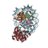 8j90C 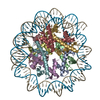 8j91C C: citing same article ( M: map data used to model this data |
|---|---|
| Similar structure data |


-Links
 PDBj
PDBj


- Assembly
| Deposited unit | 
|
|---|---|
| 1 |
|
-Components
-Protein , 4 types, 8 molecules AEBFCGDH
| #1: Protein | Mass: 15583.246 Da / Num. of mol.: 2 Source method: isolated from a genetically manipulated source Source: (gene. exp.)  #2: Protein | Mass: 11718.744 Da / Num. of mol.: 2 Source method: isolated from a genetically manipulated source Source: (gene. exp.) #3: Protein | Mass: 16280.221 Da / Num. of mol.: 2 Source method: isolated from a genetically manipulated source Source: (gene. exp.) #4: Protein | Mass: 16756.738 Da / Num. of mol.: 2 Source method: isolated from a genetically manipulated source Source: (gene. exp.) |
|---|
-DNA chain , 2 types, 2 molecules IJ
| #5: DNA chain | Mass: 51922.059 Da / Num. of mol.: 1 Source method: isolated from a genetically manipulated source Source: (gene. exp.) synthetic construct (others) / Production host: |
|---|---|
| #6: DNA chain | Mass: 52424.352 Da / Num. of mol.: 1 Source method: isolated from a genetically manipulated source Source: (gene. exp.) synthetic construct (others) / Production host: |
-Details
| Has protein modification | N |
|---|
-Experimental details
-Experiment
| Experiment | Method: ELECTRON MICROSCOPY |
|---|---|
| EM experiment | Aggregation state: PARTICLE / 3D reconstruction method: single particle reconstruction |
- Sample preparation
| Component |
| ||||||||||||||||||||||||
|---|---|---|---|---|---|---|---|---|---|---|---|---|---|---|---|---|---|---|---|---|---|---|---|---|---|
| Molecular weight | Value: 0.2 MDa / Experimental value: YES | ||||||||||||||||||||||||
| Source (natural) |
| ||||||||||||||||||||||||
| Source (recombinant) |
| ||||||||||||||||||||||||
| Buffer solution | pH: 7.5 | ||||||||||||||||||||||||
| Specimen | Embedding applied: NO / Shadowing applied: NO / Staining applied: NO / Vitrification applied: YES | ||||||||||||||||||||||||
| Vitrification | Cryogen name: ETHANE |
- Electron microscopy imaging
| Experimental equipment |  Model: Titan Krios / Image courtesy: FEI Company |
|---|---|
| Microscopy | Model: FEI TITAN KRIOS |
| Electron gun | Electron source:  FIELD EMISSION GUN / Accelerating voltage: 300 kV / Illumination mode: FLOOD BEAM FIELD EMISSION GUN / Accelerating voltage: 300 kV / Illumination mode: FLOOD BEAM |
| Electron lens | Mode: BRIGHT FIELD / Nominal defocus max: 25000 nm / Nominal defocus min: 10000 nm |
| Specimen holder | Specimen holder model: FEI TITAN KRIOS AUTOGRID HOLDER |
| Image recording | Electron dose: 59.1 e/Å2 / Film or detector model: GATAN K3 BIOQUANTUM (6k x 4k) |
| EM imaging optics | Energyfilter name: GIF Bioquantum / Energyfilter slit width: 20 eV |
- Processing
| EM software |
| ||||||||||||||||||||||||
|---|---|---|---|---|---|---|---|---|---|---|---|---|---|---|---|---|---|---|---|---|---|---|---|---|---|
| CTF correction | Type: PHASE FLIPPING AND AMPLITUDE CORRECTION | ||||||||||||||||||||||||
| 3D reconstruction | Resolution: 2.9 Å / Resolution method: FSC 0.143 CUT-OFF / Num. of particles: 196430 / Symmetry type: POINT | ||||||||||||||||||||||||
| Refine LS restraints |
|
