 Movie
Movie Controller

[English] 日本語
 Yorodumi
- PDB-8j7c: Crystal structure of triosephosphate isomerase from Leishmania or... -
+ Open data

- Basic information
| Entry | Database: PDB / ID: 8j7c | |||||||||
|---|---|---|---|---|---|---|---|---|---|---|
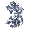
| Title | Crystal structure of triosephosphate isomerase from Leishmania orientalis at 1.88A with an arsenic ion bound at Cys57 | |||||||||
 Components Components | Triosephosphate isomerase | |||||||||
 Keywords Keywords | ISOMERASE / triosephosphate isomerase / TIM-barrel / dimer / an arsenic atom bound at Cys57 | |||||||||
| Function / homology |  Function and homology information Function and homology informationtriose-phosphate isomerase / triose-phosphate isomerase activity / glyceraldehyde-3-phosphate biosynthetic process / glycerol catabolic process / gluconeogenesis / glycolytic process / cytosol Similarity search - Function | |||||||||
| Biological species |  Leishmania orientalis (eukaryote) Leishmania orientalis (eukaryote) | |||||||||
| Method |  X-RAY DIFFRACTION / SYNCHROTRON / MOLECULAR REPLACEMENT / Resolution: 1.88 Å X-RAY DIFFRACTION / SYNCHROTRON / MOLECULAR REPLACEMENT / Resolution: 1.88 Å | |||||||||
 Authors Authors | Kuaprasert, B. / Attarataya, J. / Riangrungroj, P. / Pornthanakasem, W. / Suginta, W. / Mungthin, M. / Leelayoova, S. / Choowongkomon, K. / Leartsakulpanich, U. | |||||||||
| Funding support |  Thailand, 1items Thailand, 1items
| |||||||||
 Citation Citation | Journal: To be published Title: Leishmania orientalis triosephosphate isomerase crystal structure at 1.45 angstroms resolution and its potential specific inhibitors Authors: Kuaprasert, B. / Leartsakulpanich, U. / Riangrungroj, P. / Pornthanakasem, W. / Suginta, W. / Robinson, R. / Zhou, Y. / Mungthin, M. / Leelayoova, S. / Saehlee, S. / Kiriwan, D. / Choowongkomon, K. | |||||||||
| History |
|
- Structure visualization
| Structure viewer | Molecule: MolmilJmol/JSmol |
|---|
- Downloads & links
-Download
| PDBx/mmCIF format | 8j7c.cif.gz | 117.7 KB | Display | PDBx/mmCIF format |
|---|---|---|---|---|
| PDB format | pdb8j7c.ent.gz | 88.6 KB | Display | PDB format |
| PDBx/mmJSON format | 8j7c.json.gz | Tree view | PDBx/mmJSON format | |
| Others |  Other downloads Other downloads |
-Validation report
| Arichive directory | https://data.pdbj.org/pub/pdb/validation_reports/j7/8j7cftp://data.pdbj.org/pub/pdb/validation_reports/j7/8j7c | HTTPS FTP |
|---|
-Related structure data
-Links
 PDBj
PDBj
- Assembly
| Deposited unit | 
| |||||||||
|---|---|---|---|---|---|---|---|---|---|---|
| 1 | 
| |||||||||
| 2 | 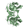
| |||||||||
| Unit cell |
| |||||||||
| Components on special symmetry positions |
|
-Components
| #1: Protein | Mass: 29244.482 Da / Num. of mol.: 2 Source method: isolated from a genetically manipulated source Source: (gene. exp.) Leishmania orientalis (eukaryote) / Gene: LSCM4_05371 / Plasmid: pET15b-LsTIMDetails (production host): lstim gene was ligated into pET15b at NdeI and BamHI sites Production host:  #2: Chemical |   Mass: 74.922 Da / Num. of mol.: 2 / Source method: obtained synthetically / Formula: As Mass: 74.922 Da / Num. of mol.: 2 / Source method: obtained synthetically / Formula: As#3: Water | ChemComp-HOH / |  Mass: 18.015 Da / Num. of mol.: 491 / Source method: isolated from a natural source / Formula: H2O Mass: 18.015 Da / Num. of mol.: 491 / Source method: isolated from a natural source / Formula: H2OHas ligand of interest | N | |
|---|
-Experimental details
-Experiment
| Experiment | Method: X-RAY DIFFRACTION / Number of used crystals: 1 |
|---|
- Sample preparation
| Crystal | Density Matthews: 2.57 Å3/Da / Density % sol: 52.12 % / Description: rod shape |
|---|---|
| Crystal grow | Temperature: 291.15 K / Method: microbatch / pH: 5.9 Details: Equal volumes (1.0 microliter) of enzyme (24.2 mg/ml in 50 mM potassium phosphate buffer, pH 8.0, containing 50 mM KCl, 5 mM DTT, and 10% glycerol) and precipitant (PEG8000 18% w/v in 0.2 mM ...Details: Equal volumes (1.0 microliter) of enzyme (24.2 mg/ml in 50 mM potassium phosphate buffer, pH 8.0, containing 50 mM KCl, 5 mM DTT, and 10% glycerol) and precipitant (PEG8000 18% w/v in 0.2 mM calcium acetate hydrate, 0.1 M and sodium cacodylate trihydrate, pH 5.9) PH range: 5.9 - 6.5 / Temp details: Incubator |
-Data collection
| Diffraction | Mean temperature: 100 K / Ambient temp details: 700 series Oxford Cryosystems / Serial crystal experiment: N |
|---|---|
| Diffraction source | Source: SYNCHROTRON / Site: SLRI / Beamline: BL7.2W / Wavelength: 1.55 Å |
| Detector | Type: MAR CCD 165 mm / Detector: CCD / Date: May 11, 2015 / Details: spherical mirror for CM and toroidal mirror for M |
| Radiation | Monochromator: SI(111) DCM / Protocol: SINGLE WAVELENGTH / Monochromatic (M) / Laue (L): M / Scattering type: x-ray |
| Radiation wavelength | Wavelength: 1.55 Å / Relative weight: 1 |
| Reflection | Resolution: 1.88→21.56 Å / Num. obs: 46160 / % possible obs: 98.6 % / Redundancy: 2.8 % / Biso Wilson estimate: 6.92 Å2 / CC1/2: 0.991 / Rmerge(I) obs: 0.082 / Rpim(I) all: 0.059 / Rrim(I) all: 0.101 / Net I/av σ(I): 7.2 / Net I/σ(I): 11.9 |
| Reflection shell | Resolution: 1.88→1.92 Å / Redundancy: 2.6 % / Rmerge(I) obs: 0.549 / Mean I/σ(I) obs: 2.4 / Num. unique obs: 2463 / CC1/2: 0.598 / Rpim(I) all: 0.399 / Rrim(I) all: 0.682 / % possible all: 81.2 |
- Processing
| Software |
| ||||||||||||||||||||||||||||||||||||||||||||||||||||||||||||||||||||||||||||||||||||||||||||||||||||||||||||||||||||||||||||||||||||||||||||||||||||||||||||||||||||||||||||||||||||||
|---|---|---|---|---|---|---|---|---|---|---|---|---|---|---|---|---|---|---|---|---|---|---|---|---|---|---|---|---|---|---|---|---|---|---|---|---|---|---|---|---|---|---|---|---|---|---|---|---|---|---|---|---|---|---|---|---|---|---|---|---|---|---|---|---|---|---|---|---|---|---|---|---|---|---|---|---|---|---|---|---|---|---|---|---|---|---|---|---|---|---|---|---|---|---|---|---|---|---|---|---|---|---|---|---|---|---|---|---|---|---|---|---|---|---|---|---|---|---|---|---|---|---|---|---|---|---|---|---|---|---|---|---|---|---|---|---|---|---|---|---|---|---|---|---|---|---|---|---|---|---|---|---|---|---|---|---|---|---|---|---|---|---|---|---|---|---|---|---|---|---|---|---|---|---|---|---|---|---|---|---|---|---|---|
| Refinement | Method to determine structure: MOLECULAR REPLACEMENT / Resolution: 1.88→19.98 Å / Cor.coef. Fo:Fc: 0.951 / Cor.coef. Fo:Fc free: 0.931 / SU B: 3.038 / SU ML: 0.087 / Cross valid method: FREE R-VALUE / ESU R: 0.131 / ESU R Free: 0.128 Stereochemistry target values: MAXIMUM LIKELIHOOD WITH PHASES Details: HYDROGENS HAVE BEEN ADDED IN THE RIDING POSITIONS
| ||||||||||||||||||||||||||||||||||||||||||||||||||||||||||||||||||||||||||||||||||||||||||||||||||||||||||||||||||||||||||||||||||||||||||||||||||||||||||||||||||||||||||||||||||||||
| Solvent computation | Ion probe radii: 0.8 Å / Shrinkage radii: 0.8 Å / VDW probe radii: 1.2 Å / Solvent model: MASK | ||||||||||||||||||||||||||||||||||||||||||||||||||||||||||||||||||||||||||||||||||||||||||||||||||||||||||||||||||||||||||||||||||||||||||||||||||||||||||||||||||||||||||||||||||||||
| Displacement parameters | Biso mean: 16.047 Å2
| ||||||||||||||||||||||||||||||||||||||||||||||||||||||||||||||||||||||||||||||||||||||||||||||||||||||||||||||||||||||||||||||||||||||||||||||||||||||||||||||||||||||||||||||||||||||
| Refinement step | Cycle: 1 / Resolution: 1.88→19.98 Å
| ||||||||||||||||||||||||||||||||||||||||||||||||||||||||||||||||||||||||||||||||||||||||||||||||||||||||||||||||||||||||||||||||||||||||||||||||||||||||||||||||||||||||||||||||||||||
| Refine LS restraints |
|