 Movie
Movie Controller

[English] 日本語
 Yorodumi
- PDB-8j3t: Complex structure of human cytomegalovirus protease and a non-cov... -
+ Open data

- Basic information
| Entry | Database: PDB / ID: 8j3t | ||||||
|---|---|---|---|---|---|---|---|
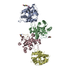
| Title | Complex structure of human cytomegalovirus protease and a non-covalent small-molecule ligand | ||||||
 Components Components | Assemblin | ||||||
 Keywords Keywords | HYDROLASE / Protease | ||||||
| Function / homology |  Function and homology information Function and homology informationassemblin / nuclear capsid assembly / viral release from host cell / host cell cytoplasm / serine-type endopeptidase activity / host cell nucleus / proteolysis / identical protein binding Similarity search - Function | ||||||
| Biological species |   Human betaherpesvirus 5 Human betaherpesvirus 5 | ||||||
| Method |  X-RAY DIFFRACTION / SYNCHROTRON / MOLECULAR REPLACEMENT / Resolution: 2.9 Å X-RAY DIFFRACTION / SYNCHROTRON / MOLECULAR REPLACEMENT / Resolution: 2.9 Å | ||||||
 Authors Authors | Yoshida, S. / Sako, Y. / Nikaido, E. / Ueda, T. / Kozono, I. / Ichihashi, Y. / Nakahashi, A. / Onishi, M. / Yamatsu, Y. / Kato, T. ...Yoshida, S. / Sako, Y. / Nikaido, E. / Ueda, T. / Kozono, I. / Ichihashi, Y. / Nakahashi, A. / Onishi, M. / Yamatsu, Y. / Kato, T. / Nishikawa, J. / Tachibana, Y. | ||||||
| Funding support | 1items
| ||||||
 Citation Citation | Journal: Acs Med.Chem.Lett. / Year: 2023 Title: Peptide-to-Small Molecule: Discovery of Non-Covalent, Active-Site Inhibitors of beta-Herpesvirus Proteases. Authors: Yoshida, S. / Sako, Y. / Nikaido, E. / Ueda, T. / Kozono, I. / Ichihashi, Y. / Nakahashi, A. / Onishi, M. / Yamatsu, Y. / Kato, T. / Nishikawa, J. / Tachibana, Y. | ||||||
| History |
|
- Structure visualization
| Structure viewer | Molecule: MolmilJmol/JSmol |
|---|
- Downloads & links
-Download
| PDBx/mmCIF format | 8j3t.cif.gz | 98.4 KB | Display | PDBx/mmCIF format |
|---|---|---|---|---|
| PDB format | pdb8j3t.ent.gz | 71.9 KB | Display | PDB format |
| PDBx/mmJSON format | 8j3t.json.gz | Tree view | PDBx/mmJSON format | |
| Others |  Other downloads Other downloads |
-Validation report
| Summary document | 8j3t_validation.pdf.gz | 1 MB | Display | wwPDB validaton report |
|---|---|---|---|---|
| Full document | 8j3t_full_validation.pdf.gz | 1 MB | Display | |
| Data in XML | 8j3t_validation.xml.gz | 18.3 KB | Display | |
| Data in CIF | 8j3t_validation.cif.gz | 23.7 KB | Display | |
| Arichive directory | https://data.pdbj.org/pub/pdb/validation_reports/j3/8j3tftp://data.pdbj.org/pub/pdb/validation_reports/j3/8j3t | HTTPS FTP |
-Related structure data
-Links
 PDBj
PDBj- Assembly
| Deposited unit | 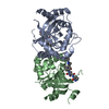
| ||||||||
|---|---|---|---|---|---|---|---|---|---|
| 1 |
| ||||||||
| Unit cell |
|
-Components
| #1: Protein | Mass: 29130.686 Da / Num. of mol.: 2 / Mutation: A143Q Source method: isolated from a genetically manipulated source Source: (gene. exp.) Human betaherpesvirus 5 / Gene: UL80 / Production host:  #2: Chemical |   Mass: 687.830 Da / Num. of mol.: 2 / Source method: obtained synthetically / Formula: C40H45N7O4 / Feature type: SUBJECT OF INVESTIGATION Mass: 687.830 Da / Num. of mol.: 2 / Source method: obtained synthetically / Formula: C40H45N7O4 / Feature type: SUBJECT OF INVESTIGATION#3: Water | ChemComp-HOH / |  Mass: 18.015 Da / Num. of mol.: 1 / Source method: isolated from a natural source / Formula: H2O Mass: 18.015 Da / Num. of mol.: 1 / Source method: isolated from a natural source / Formula: H2OHas ligand of interest | Y | |
|---|
-Experimental details
-Experiment
| Experiment | Method: X-RAY DIFFRACTION / Number of used crystals: 1 |
|---|
- Sample preparation
| Crystal | Density Matthews: 2.69 Å3/Da / Density % sol: 54.28 % |
|---|---|
| Crystal grow | Temperature: 277 K / Method: vapor diffusion, sitting drop Details: 0.05 M MES pH 5.5, 0.6 M NaCl, 0.097 M ammonium acetate, 0.005 M MgSO4 |
-Data collection
| Diffraction | Mean temperature: 100 K / Serial crystal experiment: N |
|---|---|
| Diffraction source | Source: SYNCHROTRON / Site: Photon Factory  / Beamline: BL-17A / Wavelength: 1 Å / Beamline: BL-17A / Wavelength: 1 Å |
| Detector | Type: DECTRIS EIGER X 16M / Detector: PIXEL / Date: Feb 13, 2020 |
| Radiation | Protocol: SINGLE WAVELENGTH / Monochromatic (M) / Laue (L): M / Scattering type: x-ray |
| Radiation wavelength | Wavelength: 1 Å / Relative weight: 1 |
| Reflection | Resolution: 2.8→71.96 Å / Num. obs: 16515 / % possible obs: 99.9 % / Redundancy: 13 % / CC1/2: 0.999 / Rmerge(I) obs: 0.121 / Net I/σ(I): 15.2 |
| Reflection shell | Resolution: 2.8→2.95 Å / Num. unique obs: 2345 / CC1/2: 0.959 |
- Processing
| Software |
| ||||||||||||||||||||||||||||||||||||||||||||||||||||||||||||||||||||||||||||||||||||||||||||||||||||||||||||||||||||||||||||||||||||||||||||||||||||||||||||||||||||||||||||||||||||||
|---|---|---|---|---|---|---|---|---|---|---|---|---|---|---|---|---|---|---|---|---|---|---|---|---|---|---|---|---|---|---|---|---|---|---|---|---|---|---|---|---|---|---|---|---|---|---|---|---|---|---|---|---|---|---|---|---|---|---|---|---|---|---|---|---|---|---|---|---|---|---|---|---|---|---|---|---|---|---|---|---|---|---|---|---|---|---|---|---|---|---|---|---|---|---|---|---|---|---|---|---|---|---|---|---|---|---|---|---|---|---|---|---|---|---|---|---|---|---|---|---|---|---|---|---|---|---|---|---|---|---|---|---|---|---|---|---|---|---|---|---|---|---|---|---|---|---|---|---|---|---|---|---|---|---|---|---|---|---|---|---|---|---|---|---|---|---|---|---|---|---|---|---|---|---|---|---|---|---|---|---|---|---|---|
| Refinement | Method to determine structure: MOLECULAR REPLACEMENT / Resolution: 2.9→71.96 Å / Cor.coef. Fo:Fc: 0.938 / Cor.coef. Fo:Fc free: 0.871 / SU B: 19.829 / SU ML: 0.366 / Cross valid method: THROUGHOUT / ESU R: 1.01 / ESU R Free: 0.434 / Stereochemistry target values: MAXIMUM LIKELIHOOD / Details: HYDROGENS HAVE BEEN ADDED IN THE RIDING POSITIONS
| ||||||||||||||||||||||||||||||||||||||||||||||||||||||||||||||||||||||||||||||||||||||||||||||||||||||||||||||||||||||||||||||||||||||||||||||||||||||||||||||||||||||||||||||||||||||
| Solvent computation | Ion probe radii: 0.8 Å / Shrinkage radii: 0.8 Å / VDW probe radii: 1.2 Å / Solvent model: MASK | ||||||||||||||||||||||||||||||||||||||||||||||||||||||||||||||||||||||||||||||||||||||||||||||||||||||||||||||||||||||||||||||||||||||||||||||||||||||||||||||||||||||||||||||||||||||
| Displacement parameters | Biso mean: 76.361 Å2
| ||||||||||||||||||||||||||||||||||||||||||||||||||||||||||||||||||||||||||||||||||||||||||||||||||||||||||||||||||||||||||||||||||||||||||||||||||||||||||||||||||||||||||||||||||||||
| Refinement step | Cycle: 1 / Resolution: 2.9→71.96 Å
| ||||||||||||||||||||||||||||||||||||||||||||||||||||||||||||||||||||||||||||||||||||||||||||||||||||||||||||||||||||||||||||||||||||||||||||||||||||||||||||||||||||||||||||||||||||||
| Refine LS restraints |
|