 Movie
Movie Controller

[English] 日本語
 Yorodumi
- PDB-8j2c: Structure of the C-terminal subenzyme of the malonyl-CoA reductas... -
+ Open data

- Basic information
| Entry | Database: PDB / ID: 8j2c | ||||||
|---|---|---|---|---|---|---|---|
| Title | Structure of the C-terminal subenzyme of the malonyl-CoA reductase from Chloroflexus aurantiacus, mutant N940V/K1106W/S1114R | ||||||
 Components Components | Short-chain dehydrogenase/reductase SDR | ||||||
 Keywords Keywords | OXIDOREDUCTASE / 3-hydroxypropionate(3-HP) / malonyl-CoA reductase / CO2 fixation / short-chain dehydrogenase/reductase (SDR) / Chloroflexus aurantiacus | ||||||
| Function / homology |  Function and homology information Function and homology informationfatty acid elongation / oxidoreductase activity, acting on the CH-OH group of donors, NAD or NADP as acceptor / nucleotide binding / metal ion binding Similarity search - Function | ||||||
| Biological species |   Chloroflexus aurantiacus (bacteria) Chloroflexus aurantiacus (bacteria) | ||||||
| Method |  X-RAY DIFFRACTION / SYNCHROTRON / MOLECULAR REPLACEMENT / Resolution: 1.7 Å X-RAY DIFFRACTION / SYNCHROTRON / MOLECULAR REPLACEMENT / Resolution: 1.7 Å | ||||||
 Authors Authors | Ma, Q. / Liu, C. | ||||||
| Funding support |  China, 1items China, 1items
| ||||||
 Citation Citation | Journal: To Be Published Title: Structures of the C-terminal subenzyme of the malonyl-CoA reductase from Chloroflexus aurantiacus Authors: Ma, Q. / Liu, C. | ||||||
| History |
|
- Structure visualization
| Structure viewer | Molecule: MolmilJmol/JSmol |
|---|
- Downloads & links
-Download
| PDBx/mmCIF format | 8j2c.cif.gz | 270.2 KB | Display | PDBx/mmCIF format |
|---|---|---|---|---|
| PDB format | pdb8j2c.ent.gz | 213.9 KB | Display | PDB format |
| PDBx/mmJSON format | 8j2c.json.gz | Tree view | PDBx/mmJSON format | |
| Others |  Other downloads Other downloads |
-Validation report
| Summary document | 8j2c_validation.pdf.gz | 444.3 KB | Display | wwPDB validaton report |
|---|---|---|---|---|
| Full document | 8j2c_full_validation.pdf.gz | 444.6 KB | Display | |
| Data in XML | 8j2c_validation.xml.gz | 27.5 KB | Display | |
| Data in CIF | 8j2c_validation.cif.gz | 42.7 KB | Display | |
| Arichive directory | https://data.pdbj.org/pub/pdb/validation_reports/j2/8j2cftp://data.pdbj.org/pub/pdb/validation_reports/j2/8j2c | HTTPS FTP |
-Related structure data

-Links
 PDBj
PDBj


- Assembly
| Deposited unit | 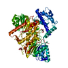
| ||||||||
|---|---|---|---|---|---|---|---|---|---|
| 1 | 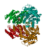
| ||||||||
| 2 |
| ||||||||
| Unit cell |
| ||||||||
| Components on special symmetry positions |
|
-Components
| #1: Protein | Mass: 73764.039 Da / Num. of mol.: 1 / Mutation: N940V/K1106W/S1114R Source method: isolated from a genetically manipulated source Source: (gene. exp.) Chloroflexus aurantiacus (strain ATCC 29366 / DSM 635 / J-10-fl) (bacteria)Gene: Caur_2614 / Production host: |
|---|---|
| #2: Chemical | ChemComp-TLA /   Mass: 150.087 Da / Num. of mol.: 1 / Source method: obtained synthetically / Formula: C4H6O6 Mass: 150.087 Da / Num. of mol.: 1 / Source method: obtained synthetically / Formula: C4H6O6 |
| #3: Chemical | ChemComp-GOL /   Mass: 92.094 Da / Num. of mol.: 1 / Source method: obtained synthetically / Formula: C3H8O3 Mass: 92.094 Da / Num. of mol.: 1 / Source method: obtained synthetically / Formula: C3H8O3 |
| #4: Water | ChemComp-HOH /  Mass: 18.015 Da / Num. of mol.: 523 / Source method: isolated from a natural source / Formula: H2O Mass: 18.015 Da / Num. of mol.: 523 / Source method: isolated from a natural source / Formula: H2O |
| Has ligand of interest | N |
-Experimental details
-Experiment
| Experiment | Method: X-RAY DIFFRACTION / Number of used crystals: 1 |
|---|
- Sample preparation
| Crystal | Density Matthews: 2.93 Å3/Da / Density % sol: 58.07 % |
|---|---|
| Crystal grow | Temperature: 293 K / Method: vapor diffusion, sitting drop Details: The protein was crystallized in drops containing 1.5 ul protein solution (5 mg/ml) and 1.5 ul reservoir solution (1.1 M ammonium tartrate pH 7.0) |
-Data collection
| Diffraction | Mean temperature: 100 K / Serial crystal experiment: N |
|---|---|
| Diffraction source | Source: SYNCHROTRON / Site: SSRF / Beamline: BL19U1 / Wavelength: 0.97774 Å |
| Detector | Type: DECTRIS PILATUS3 6M / Detector: PIXEL / Date: Jun 23, 2017 |
| Radiation | Protocol: SINGLE WAVELENGTH / Monochromatic (M) / Laue (L): M / Scattering type: x-ray |
| Radiation wavelength | Wavelength: 0.97774 Å / Relative weight: 1 |
| Reflection | Resolution: 1.697→73.265 Å / Num. obs: 91959 / % possible obs: 98.2 % / Redundancy: 6.7 % / CC1/2: 0.999 / Rpim(I) all: 0.024 / Rrim(I) all: 0.063 / Rsym value: 0.058 / Net I/σ(I): 17.3 |
| Reflection shell | Resolution: 1.697→1.702 Å / Redundancy: 5.7 % / Mean I/σ(I) obs: 2.2 / Num. unique obs: 734 / CC1/2: 0.815 / Rpim(I) all: 0.277 / Rrim(I) all: 0.674 / Rsym value: 0.612 / % possible all: 76.1 |
- Processing
| Software |
| ||||||||||||||||||||||||||||||||||||||||||||||||||||||||||||||||||||||||||||||||||||||||||||||||||||||||||||||||||
|---|---|---|---|---|---|---|---|---|---|---|---|---|---|---|---|---|---|---|---|---|---|---|---|---|---|---|---|---|---|---|---|---|---|---|---|---|---|---|---|---|---|---|---|---|---|---|---|---|---|---|---|---|---|---|---|---|---|---|---|---|---|---|---|---|---|---|---|---|---|---|---|---|---|---|---|---|---|---|---|---|---|---|---|---|---|---|---|---|---|---|---|---|---|---|---|---|---|---|---|---|---|---|---|---|---|---|---|---|---|---|---|---|---|---|---|
| Refinement | Method to determine structure: MOLECULAR REPLACEMENT / Resolution: 1.7→73.265 Å / Cor.coef. Fo:Fc: 0.964 / Cor.coef. Fo:Fc free: 0.961 / SU R Cruickshank DPI: 0.082 / Cross valid method: THROUGHOUT / σ(F): 0 / SU R Blow DPI: 0.083 / SU Rfree Blow DPI: 0.08 / SU Rfree Cruickshank DPI: 0.079
| ||||||||||||||||||||||||||||||||||||||||||||||||||||||||||||||||||||||||||||||||||||||||||||||||||||||||||||||||||
| Displacement parameters | Biso mean: 30.81 Å2
| ||||||||||||||||||||||||||||||||||||||||||||||||||||||||||||||||||||||||||||||||||||||||||||||||||||||||||||||||||
| Refine analyze | Luzzati coordinate error obs: 0.19 Å | ||||||||||||||||||||||||||||||||||||||||||||||||||||||||||||||||||||||||||||||||||||||||||||||||||||||||||||||||||
| Refinement step | Cycle: 1 / Resolution: 1.7→73.265 Å
| ||||||||||||||||||||||||||||||||||||||||||||||||||||||||||||||||||||||||||||||||||||||||||||||||||||||||||||||||||
| Refine LS restraints |
| ||||||||||||||||||||||||||||||||||||||||||||||||||||||||||||||||||||||||||||||||||||||||||||||||||||||||||||||||||
| LS refinement shell | Resolution: 1.7→1.74 Å / Rfactor Rfree error: 0 / Total num. of bins used: 20
| ||||||||||||||||||||||||||||||||||||||||||||||||||||||||||||||||||||||||||||||||||||||||||||||||||||||||||||||||||
| Refinement TLS params. | Method: refined / Origin x: -33.728 Å / Origin y: 6.8011 Å / Origin z: -14.9176 Å
| ||||||||||||||||||||||||||||||||||||||||||||||||||||||||||||||||||||||||||||||||||||||||||||||||||||||||||||||||||
| Refinement TLS group | Selection details: { A|562 - A|1219 } |
