regulation of calcium ion transport into cytosol / ubiquitin-like protein ligase activity / negative regulation of translation in response to oxidative stress / positive regulation of protein localization to chromosome, telomeric region / suppression of viral release by host / PML body organization / SUMO binding / positive regulation of apoptotic process involved in mammary gland involution / SMAD protein signal transduction / fibroblast migration ...regulation of calcium ion transport into cytosol / ubiquitin-like protein ligase activity / negative regulation of translation in response to oxidative stress / positive regulation of protein localization to chromosome, telomeric region / suppression of viral release by host / PML body organization / SUMO binding / positive regulation of apoptotic process involved in mammary gland involution / SMAD protein signal transduction / fibroblast migration / myeloid cell differentiation / protein-containing complex localization / maintenance of protein location in nucleus / endoplasmic reticulum calcium ion homeostasis / regulation of double-strand break repair / branching involved in mammary gland duct morphogenesis / oncogene-induced cell senescence / negative regulation of mitotic cell cycle / Transferases; Acyltransferases; Aminoacyltransferases / SUMO transferase activity / Regulation of RUNX1 Expression and Activity / positive regulation of extrinsic apoptotic signaling pathway / cobalt ion binding / intrinsic apoptotic signaling pathway in response to oxidative stress / negative regulation of interleukin-1 beta production / entrainment of circadian clock by photoperiod / SUMOylation of ubiquitinylation proteins / SMAD binding / detection of maltose stimulus / maltose transport complex / negative regulation of telomere maintenance via telomerase / carbohydrate transport / positive regulation of telomere maintenance / protein sumoylation / intrinsic apoptotic signaling pathway in response to endoplasmic reticulum stress / carbohydrate transmembrane transporter activity / maltose binding / intrinsic apoptotic signaling pathway in response to DNA damage by p53 class mediator / cell fate commitment / negative regulation of ubiquitin-dependent protein catabolic process / maltose transport / maltodextrin transmembrane transport / positive regulation of signal transduction by p53 class mediator / regulation of cell adhesion / SUMOylation of DNA damage response and repair proteins / protein targeting / response to UV / retinoic acid receptor signaling pathway / ATP-binding cassette (ABC) transporter complex, substrate-binding subunit-containing / Regulation of TP53 Activity through Acetylation / extrinsic apoptotic signaling pathway / positive regulation of defense response to virus by host / response to cytokine / transforming growth factor beta receptor signaling pathway / cellular response to interleukin-4 / ATP-binding cassette (ABC) transporter complex / Regulation of PTEN localization / negative regulation of angiogenesis / cellular response to leukemia inhibitory factor / response to gamma radiation / cell chemotaxis / DNA damage response, signal transduction by p53 class mediator / circadian regulation of gene expression / negative regulation of cell growth / regulation of circadian rhythm / PML body / nuclear matrix / positive regulation of fibroblast proliferation / Transcriptional regulation of granulopoiesis / intrinsic apoptotic signaling pathway in response to DNA damage / Interferon gamma signaling / protein import into nucleus / HCMV Early Events / cellular senescence / outer membrane-bounded periplasmic space / protein-containing complex assembly / early endosome membrane / nuclear membrane / molecular adaptor activity / proteasome-mediated ubiquitin-dependent protein catabolic process / response to hypoxia / transcription coactivator activity / chromosome, telomeric region / periplasmic space / regulation of cell cycle / protein stabilization / chromatin remodeling / protein heterodimerization activity / negative regulation of cell population proliferation / innate immune response / negative regulation of DNA-templated transcription / apoptotic process / DNA damage response / ubiquitin protein ligase binding / regulation of DNA-templated transcription / endoplasmic reticulum membrane / nucleolus / protein homodimerization activity / DNA binding / zinc ion binding Similarity search - Function
Protein of unknown function DUF3583 / PML-like, coiled-coil / PML, C-terminal domain / : / ANCHR-like B-box zinc-binding domain / B-Box-type zinc finger / B-box-type zinc finger / Zinc finger B-box type profile. / Zinc finger, C3HC4 RING-type / Zinc finger, C3HC4 type (RING finger) ...Protein of unknown function DUF3583 / PML-like, coiled-coil / PML, C-terminal domain / : / ANCHR-like B-box zinc-binding domain / B-Box-type zinc finger / B-box-type zinc finger / Zinc finger B-box type profile. / Zinc finger, C3HC4 RING-type / Zinc finger, C3HC4 type (RING finger) / Maltose/Cyclodextrin ABC transporter, substrate-binding protein / Solute-binding family 1, conserved site / Bacterial extracellular solute-binding proteins, family 1 signature. / Bacterial extracellular solute-binding protein / Bacterial extracellular solute-binding protein / Zinc finger, RING-type, conserved site / Zinc finger RING-type signature. / Ring finger / Zinc finger RING-type profile. / Zinc finger, RING-type / Zinc finger, RING/FYVE/PHD-type Similarity search - Domain/homology
In the structure databanks used in Yorodumi, some data are registered as the other names, "COVID-19 virus" and "2019-nCoV". Here are the details of the virus and the list of structure data.
Jan 31, 2019. EMDB accession codes are about to change! (news from PDBe EMDB page)
EMDB accession codes are about to change! (news from PDBe EMDB page)
The allocation of 4 digits for EMDB accession codes will soon come to an end. Whilst these codes will remain in use, new EMDB accession codes will include an additional digit and will expand incrementally as the available range of codes is exhausted. The current 4-digit format prefixed with “EMD-” (i.e. EMD-XXXX) will advance to a 5-digit format (i.e. EMD-XXXXX), and so on. It is currently estimated that the 4-digit codes will be depleted around Spring 2019, at which point the 5-digit format will come into force.
The EM Navigator/Yorodumi systems omit the EMD- prefix.
Related info.:Q: What is EMD? / ID/Accession-code notation in Yorodumi/EM Navigator
Yorodumi is a browser for structure data from EMDB, PDB, SASBDB, etc.
This page is also the successor to EM Navigator detail page, and also detail information page/front-end page for Omokage search.
The word "yorodu" (or yorozu) is an old Japanese word meaning "ten thousand". "mi" (miru) is to see.
Related info.:EMDB / PDB / SASBDB / Comparison of 3 databanks / Yorodumi Search / Aug 31, 2016. New EM Navigator & Yorodumi / Yorodumi Papers / Jmol/JSmol / Function and homology information / Changes in new EM Navigator and Yorodumi
 Movie
Movie Controller


 Open data
 Basic information
 Components
 Keywords
 Function and homology information
 Homo sapiens (human)
 X-RAY DIFFRACTION /
  Authors
 China, 1items
  Citation
 Structure visualization
 Downloads & links
 Other downloads
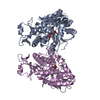
 PDBj
PDBj


 Assembly
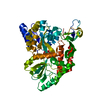

 Mass: 65.409 Da / Num. of mol.: 2 / Source method: isolated from a natural source / Formula: Zn / Feature type: SUBJECT OF INVESTIGATION
 Mass: 18.015 Da / Num. of mol.: 42 / Source method: isolated from a natural source / Formula: H2O
 Sample preparation
 Processing