 Movie
Movie Controller


+ Open data

- Basic information
| Entry | Database: PDB / ID: 8j0n | ||||||
|---|---|---|---|---|---|---|---|
| Title | cryo-EM structure of human EMC | ||||||
 Components Components | (ER membrane protein complex subunit ...) x 9 | ||||||
 Keywords Keywords | MEMBRANE PROTEIN / ER membrane protein complex | ||||||
| Function / homology |  Function and homology information Function and homology informationextrinsic component of endoplasmic reticulum membrane / EMC complex / : / omegasome membrane / protein insertion into ER membrane by stop-transfer membrane-anchor sequence / magnesium ion transport / tail-anchored membrane protein insertion into ER membrane / Miscellaneous transport and binding events / cobalt ion transmembrane transporter activity / ferrous iron transmembrane transporter activity ...extrinsic component of endoplasmic reticulum membrane / EMC complex / : / omegasome membrane / protein insertion into ER membrane by stop-transfer membrane-anchor sequence / magnesium ion transport / tail-anchored membrane protein insertion into ER membrane / Miscellaneous transport and binding events / cobalt ion transmembrane transporter activity / ferrous iron transmembrane transporter activity / copper ion transport / magnesium ion transmembrane transporter activity / RHOA GTPase cycle / autophagosome assembly / positive regulation of endothelial cell proliferation / positive regulation of endothelial cell migration / positive regulation of angiogenesis / carbohydrate binding / early endosome membrane / angiogenesis / early endosome / Golgi membrane / apoptotic process / endoplasmic reticulum membrane / endoplasmic reticulum / Golgi apparatus / protein-containing complex / extracellular region / membrane / plasma membrane / cytoplasm Similarity search - Function | ||||||
| Biological species |  Homo sapiens (human) Homo sapiens (human) | ||||||
| Method | ELECTRON MICROSCOPY / single particle reconstruction / cryo EM / Resolution: 3.47 Å | ||||||
 Authors Authors | Li, M. / Zhang, C. / Wu, J. / Lei, M. | ||||||
| Funding support |  China, 1items China, 1items
| ||||||
 Citation Citation | Journal: Aging (Albany NY) / Year: 2024 Title: Structural insights into human EMC and its interaction with VDAC. Authors: Mingyue Li / Chunli Zhang / Yuntao Xu / Shaobai Li / Chenhui Huang / Jian Wu / Ming Lei / Abstract: The endoplasmic reticulum (ER) membrane protein complex (EMC) is a conserved, multi-subunit complex acting as an insertase at the ER membrane. Growing evidence shows that the EMC is also involved in ...The endoplasmic reticulum (ER) membrane protein complex (EMC) is a conserved, multi-subunit complex acting as an insertase at the ER membrane. Growing evidence shows that the EMC is also involved in stabilizing and trafficking membrane proteins. However, the structural basis and regulation of its multifunctionality remain elusive. Here, we report cryo-electron microscopy structures of human EMC in apo- and voltage-dependent anion channel (VDAC)-bound states at resolutions of 3.47 Å and 3.32 Å, respectively. We discovered a specific interaction between VDAC proteins and the EMC at mitochondria-ER contact sites, which is conserved from yeast to humans. Moreover, we identified a gating plug located inside the EMC hydrophilic vestibule, the substrate-binding pocket for client insertion. Conformation changes of this gating plug during the apo-to-VDAC-bound transition reveal that the EMC unlikely acts as an insertase in the VDAC1-bound state. Based on the data analysis, the gating plug may regulate EMC functions by modifying the hydrophilic vestibule in different states. Our discovery offers valuable insights into the structural basis of EMC's multifunctionality. | ||||||
| History |
|
- Structure visualization
| Structure viewer | Molecule: MolmilJmol/JSmol |
|---|
- Downloads & links
-Download
| PDBx/mmCIF format | 8j0n.cif.gz | 472.1 KB | Display | PDBx/mmCIF format |
|---|---|---|---|---|
| PDB format | pdb8j0n.ent.gz | 377.7 KB | Display | PDB format |
| PDBx/mmJSON format | 8j0n.json.gz | Tree view | PDBx/mmJSON format | |
| Others |  Other downloads Other downloads |
-Validation report
| Arichive directory | https://data.pdbj.org/pub/pdb/validation_reports/j0/8j0nftp://data.pdbj.org/pub/pdb/validation_reports/j0/8j0n | HTTPS FTP |
|---|
-Related structure data
| Related structure data |  35906MC 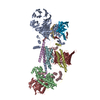 8j0oC M: map data used to model this data C: citing same article ( |
|---|---|
| Similar structure data |

-Links
 PDBj
PDBj


- Assembly
| Deposited unit | 
|
|---|---|
| 1 |
|
-Components
-ER membrane protein complex subunit ... , 9 types, 9 molecules ABCDEFGHJ
| #1: Protein | Mass: 111886.141 Da / Num. of mol.: 1 / Source method: isolated from a natural source / Source: (natural) Homo sapiens (human) / References: UniProt: Q8N766 |
|---|---|
| #2: Protein | Mass: 34882.531 Da / Num. of mol.: 1 / Source method: isolated from a natural source / Source: (natural) Homo sapiens (human) / References: UniProt: Q15006 |
| #3: Protein | Mass: 29981.924 Da / Num. of mol.: 1 / Source method: isolated from a natural source / Source: (natural) Homo sapiens (human) / References: UniProt: Q9P0I2 |
| #4: Protein | Mass: 20104.572 Da / Num. of mol.: 1 / Source method: isolated from a natural source / Source: (natural) Homo sapiens (human) / References: UniProt: Q5J8M3 |
| #5: Protein | Mass: 14706.786 Da / Num. of mol.: 1 / Source method: isolated from a natural source / Source: (natural) Homo sapiens (human) / References: UniProt: Q8N4V1 |
| #6: Protein | Mass: 12029.248 Da / Num. of mol.: 1 / Source method: isolated from a natural source / Source: (natural) Homo sapiens (human) / References: UniProt: Q9BV81 |
| #7: Protein | Mass: 26501.586 Da / Num. of mol.: 1 / Source method: isolated from a natural source / Source: (natural) Homo sapiens (human) / References: UniProt: Q9NPA0 |
| #8: Protein | Mass: 23807.076 Da / Num. of mol.: 1 / Source method: isolated from a natural source / Source: (natural) Homo sapiens (human) / References: UniProt: O43402 |
| #9: Protein | Mass: 21781.484 Da / Num. of mol.: 1 / Source method: isolated from a natural source / Source: (natural) Homo sapiens (human) / References: UniProt: Q5UCC4 |
-Sugars , 1 types, 3 molecules
| #10: Polysaccharide | Source method: isolated from a genetically manipulated source |
|---|
-Details
| Has ligand of interest | N |
|---|---|
| Has protein modification | Y |
-Experimental details
-Experiment
| Experiment | Method: ELECTRON MICROSCOPY |
|---|---|
| EM experiment | Aggregation state: PARTICLE / 3D reconstruction method: single particle reconstruction |
- Sample preparation
| Component | Name: EMC / Type: COMPLEX / Entity ID: #1-#9 / Source: NATURAL |
|---|---|
| Source (natural) | Organism: Homo sapiens (human) |
| Buffer solution | pH: 7.5 |
| Specimen | Embedding applied: NO / Shadowing applied: NO / Staining applied: NO / Vitrification applied: YES |
| Vitrification | Cryogen name: ETHANE |
- Electron microscopy imaging
| Experimental equipment |  Model: Titan Krios / Image courtesy: FEI Company |
|---|---|
| Microscopy | Model: FEI TITAN KRIOS |
| Electron gun | Electron source:  FIELD EMISSION GUN / Accelerating voltage: 300 kV / Illumination mode: FLOOD BEAM FIELD EMISSION GUN / Accelerating voltage: 300 kV / Illumination mode: FLOOD BEAM |
| Electron lens | Mode: BRIGHT FIELD / Nominal defocus max: 2500 nm / Nominal defocus min: 1200 nm |
| Image recording | Electron dose: 50 e/Å2 / Film or detector model: GATAN K3 (6k x 4k) |
- Processing
| EM software | Name: PHENIX / Version: 1.20.1_4487: / Category: model refinement | ||||||||||||||||||||||||
|---|---|---|---|---|---|---|---|---|---|---|---|---|---|---|---|---|---|---|---|---|---|---|---|---|---|
| CTF correction | Type: PHASE FLIPPING AND AMPLITUDE CORRECTION | ||||||||||||||||||||||||
| 3D reconstruction | Resolution: 3.47 Å / Resolution method: FSC 0.143 CUT-OFF / Num. of particles: 693381 / Symmetry type: POINT | ||||||||||||||||||||||||
| Refine LS restraints |
|
